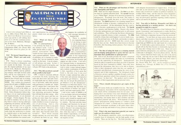
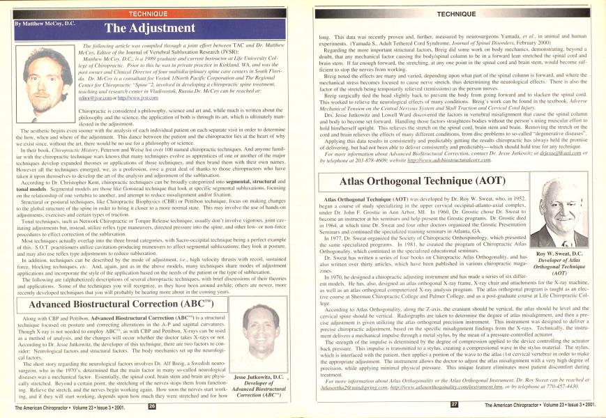
The following article was compiled through a joint effort between TAC and Dr. Matthew McCoy, Editor of the Journal of Vertebral Subluxation Research (JVSR): Matthew McCox. D.C., is a I9SV graduate and current Instructor at Life University College oj Chiropractic. Prior to this he was in private practice in Kirkland. W.A. and was the past owner and Clinical Director of four multidisciplinarx spine care centers in South Florida. Dr. McCox is a consultant for \'oslok I/North Pacific Corporation and The Regional Center for Chiropractic "Spine"2. involved in developing a chiropractic spine treatment, leaching and research center in Vladivostok. Russia.Dr. McCox can he reached at: editortg'jvsr.com or hltp:/Avww.jvsr.com Chiropractic is considered a philosophy, science and art and. while much is written about the philosophy and the science, the application ol both is through its art, which is ultimately manifested in the adjustment. The aesthetic begins even sooner with the analysis of each individual patient on each separate visit in order to determine the how. when and where of the adjustment. This dance between the patient and the chiropractor lies at the heart of why we exist since, without the art. there would be no use for a philosophy or science. In their book. Cliiiopiaclic History. Peterson and Weise list over 100 named chiropractic techniques. And anyone familiar with the chiropractic technique wars knows that many techniques evolve as apprentices of one or another of the major techniques develop expanded theories or applications of those techniques, and then brand them with their own names. However all the techniques emerged, we. as a profession, owe a great deal of thanks to those chiropractors who have taken it upon themselves to develop the art of the analysis and adjustment of the suhluxation. According to Dr. Christopher Kent, chiropractic techniques can be broadly categorized into sejjim'iital. structural and tonal models. Segmental models are those like Gonstead technique that look at specific scgmental subluxalions. focusing on the relationship of one vertebra to another, and attempt to reduce misalignment and/or fixation. Structural or postural techniques, like Chiropractic Biophysics (CBR) or Peltibon technique, focus on making changes to the global structure of the spine in order to bring it closer to a more normal state. This may involve the use of hands-on adjustments, exercises and certain types of traction. Tonal techniques, such as Network Chiropractic or Torque Release technique, usually don't involve vigorous, joint cav-itating adjustments but. instead, utilize reflex type maneuvers, directed pressure into the spine, and other low- or non-force procedures to effect correction of the subluxalion. Most techniques actually overlap into the three broad categories, with Sacro-occipital technique being a perfect example of this. S.O.T. practitioners utilize cavitation-producing maneuvers to affect segmental subluxations: they look at posture, and may also use reflex type adjustments to reduce subluxation. In addition, techniques can be described by the mode of adjustment, i.e.. high velocity thrusts with recoil, sustained force, blocking techniques, etc. And. again, just as in the above models, many techniques share modes of adjustment applications and incorporate the style of the application based on the needs of the patient or the type of subluxalion. The following are (alphabetized) descriptions of several chiropractic techniques, with brief discussions of their theories and applications. Some of the techniques you will recognize, as they have been around awhile: others are newer, more recently developed techniques that you will probably be hearing more about in the coming years. Advanced Biostructural Correction (ABC') Along with C'BP and Peltibon. Advanced Biostructural Correction (ABC") is a structural technique focused on posture and correcting alterations in the A-P and sagittal curvatures. Though X-ray is not needed to employ ABC", as with CBP and Pettibon, X-rays can be used as a method of analysis, and the changes will occur whether the doctor takes X-rays or not. According to Dr. Jesse Julkowitz. the developer of this technique, there are two factors to consider: Neurological factors and structural factors. The body mechanics set up the neurological factors. The short story regarding the neurological factors involves Dr. All' Breig, a Swedish neuro-surgeon. who in the 1970\s. determined that the main factor in many so-called neurological diseases was a mechanical factor. Essentially, the spinal cord, brain stem and brain are physically stretched. Beyond a certain point, the stretching of the nerves stops them from functioning. Relieve the stretch, and the nerves begin working again. How soon the nerves start working, and if they will start working, depends upon how much they were stretched and for how long. This data was recently proven and. further, measured by neurosurgeons Yamada. el at., in animal and human experiments. (Yamada S.. Adult Tethered Cord Syndrome. Journal of Spinal Disorders. February 2000) Regarding the more important structural factors. Breig did some work on body mechanics, demonstrating, beyond a doubt, that any mechanical factor causing the body/spinal column to be in a forward lean stretched the spinal cord and brain stem. If far enough forward, the stretching, at any one point in the spinal cord and brain stem, would become sufficient to stop the nerves from working. Breig noted the effects are many and varied, depending upon what part of the spinal column is forward, and where the mechanical stress becomes focused to cause nerve stretch, thus determining the neurological effects. There is also the factor of the stretch being temporarily relieved (remissions) as the person moves. Breig surgically tied the head slightly back to prevent the body from going forward and to slacken the spinal cord. This worked to relieve the neurological effects of many conditions. Breig's work can be found in the textbook. Adverse Mechanical Tension on the Central Nervous System and Skull Traction and Cervical Cord Injury. Drs. Jesse Jutkowitz and Lowell Ward discovered the factors in vertebral misalignment that cause the spinal column and body to become set forward. Handling those factors straightens bodies without the person's using muscular effort to hold him/herself upright. This relieves the stretch on the spinal cord, brain stem and brain. Removing the stretch on the cord and brain relieves the effects of main different conditions, from disc problems to so-called "degenerative diseases". Applying this data results in consistently and predictably getting the results chiropractic has always held the promise of delivering, but had not been able to deliver consistently and predictably—which should hold true for any technique. For more information about Advanced BioStructural Correction, contact Dr. Jesse Jutkowitz at driesseii @ aol.com or by telephone at 203-878-4609: website htip://w\\w.advbiosiructuralcorr.coni. Atlas Orthogonal Technique (AOT) Atlas Orthogonal Technique (AOT) was developed by Dr. Roy W. Sweat, who. in 1952. began a course of study specializing in the upper cervical occipital-atlanto-axial complex, under Dr. John F. Grostic in Ann Arbor. MI. In I960. Dr. Grostic chose Dr. Sweat to become an instructor at his seminars and help present the Grostic programs. Dr. Grostic died in 1964. at which time Dr. Sweat and four other doctors organized the Grostic Presentation Seminars and continued the specialized training seminars in Atlanta. GA. In 1977. Dr. Sweat organized the SocietN of Chiropractic Orthospinology. which presented the same specialized programs. In 1981. he created the program of Chiropractic Atlas Orthogonality, which continued in the specialized educational seminars. Dr. Sweat has written a series of four books on Chiropractic Atlas Orthogonality, and has also written over thirty articles, which have been published in various chiropractic magazines. In 1970. he designed a chiropractic adjusting instrument and has made a series of six differ- cut models. He has. also, designed an atlas orthogonal X-ray frame. X-ray chair and attachments for the X-ray machine. as well as an atlas orthogonal computerized X-ray analysis program. The atlas orthogonal program is taught as an elective course at Sherman Chiropractic College and Palmer College, and as a post-graduate course at Life Chiropractic College. According to Atlas Orthogonality, along the Z-a\is. the cranium should be vertical, the atlas should be level and the cervical spine should be vertical. Radiographs are taken to determine the degree of atlas misalignment, and then a precise adjustment is given utilizing the atlas orthogonal precision instrument. This instrument was designed to deliver a precise chiropractic adjustment, based on the specific misalignment findings from the X-rays. Technically, the instrument delivers a mechanical impulse through a metal stylus, by the mean of a pressure-controlled actuator. The strength of the impulse is determined by the degree of compression applied to the device controlling the actuator back pressure. This impulse is transmitted to a stylus, creating a compressional wave in the stylus material. The stylus, which is interfaced with the patient, then applies a portion of the wave to the atlas (1st cervical vertebra) in order to make the appropriate adjustment. The instrument allows the doctor to adjust the atlas misalignment with a very high degree of precision, while applying minimal physical pressure. This unique feature eliminates most patient discomfort during treatment. For more information about Atlas Orthogonality or the Atlas Orthogonal Instrument. Dr. Roy Sweat can be reached at Atlasoriho2<i> inintlsprinu.com. http://\\\\w.atlasorthovonalit\.com/instrument.htm, or by telephone at 770-457-4430. Bio-Geometric Integration (BGI) Bio-Geometric Integration (BGI) was developed by Dr. Sue Brown, and is a conceptual understanding that enhances the chiropractor's knowledge of the human body. BGI provides an understanding of the innate geometry of (lie body and the force dynamics surrounding the creation and the release ol suhluxations. The philosophy, science, and art of chiropractic are examined from a post-Newtonian point of view, providing the opportunity to express and understand chiropractic in accord with contemporary science. Through understanding the innate geometry of the body, the chiropractor is more able to efficiently and gently release the subluxation. and assess the effectiveness of the adjustment. The geometric understanding of the body also serves to bridge the gap between the many techniques o( chiropractic, by providing a common language and understanding from which to converse. Bio-Geometric Integration empowers chiropractors with, not only the "how" of adjusting, but the "why", blending perfectly the philosophical concepts of chiropractic with the science of quantum physics. It deals with an understanding of the innate geometry of the body and the force dynamics surrounding the creation and release of subluxations. In BGI, the cause of subluxations is explained as "an unintegrated experience of new force frequencies". If the body cannot fully integrate and dissipate the forces to which it is exposed, it will then store the energy contained in these forces. This energy/force is stored as tension in the physical structures of the body, with the muscular, skeletal and liga-mentous systems being the most commonly involved. Tension in these systems causes tension/compression on the nervous system and. thus, subluxations are formed. Release of the stored force/energy/tone will release the tension and. thus, the suhluxation. BGI is currently working on a research project involving the investigation of long-term changes in the level of ego development of people receiving chiropractic care. BGI seminars are taught throughout the United States. For more information ahoul Bio-Geometric Integration, contact Dr. Sue Brown at Bi;iscminars({?aol.coin. or by telephone at 630-.690-6080. Chiropractic Biophysics Chiropractic Biophysics(CBP)® was developed by Dr. Don Harrison, who. along with several others, lias gone to great lengths to research and document the validity of CBP technique. As a result. 48 papers have been published/accepted in peer reviewed journals to date, with 4 more in the review process. These have provided a strong argument for the existence of a normal spinal model. The idea that a normal spine should be straight in the A-P and curved in the sagittal is taken for granted by most chiropractors. The fact that this had to be proven seems strange, at first: but. for example, many contend that curvatures in the A-P plane are normal variants, or compensatory, while also asserting that loss or even reversal of a cervical lordosis is also a normal variant, requiring no correction. These sorts of contentions are exactly where Harrison, el ai. have directed their research efforts, and they have quite a lot to show for it. Finding its roots in upper cervical chiropractic of the I93()'s with Dr. A. A. Wernsing. through B.J. Palmer. Grostic and Peltibon. CBP® developed out of the common theme that there is a normal structural orientation it) the spine. With that in mind, they felt chiropractic should attempt to bring the patient in line with this normal model in order to remove interference to the functioning of the nervous system. Chiropractic Biophysics is a full spine technique, placing emphasis on three-dimensional postural distortions and radi-ographic images. CBP® goes beyond simple postural examination, realizing that there are. in fact. 728 possible postural permutations of the skull to the thoracic cage. 728 possibilities of the thoracic cage to the pelvis, and 242 possible pelvic postures. Utilizing a systematic analysis of posture, based on the Cartesian coordinate system, the patient is then adjusted in a mirror image fashion, utilizing a drop table or a hand held instrument in order to effect postural and structural correction. In addition to the adjustment, specific mirror image exercises may be given to help reduce the distortions. If there is a loss of a sagittal curvature, specific extension traction procedures are used to restore the curves. In CBP® technique, sithlu.xation and misalignment are synonymous: so. palpation of the static or motion variety to assess joint play is not utilized in CBP®. and neither are leg length checks. The primary diagnostic tools of the CBP practitioner are postural assessment and X-ray analysis. Great care is taken in CBP to make sure that there is no distortion in the X-ray images, since these films will be used to determine what is wrong and what will be done for the patient. Alignment of the X-ray frame, tube. etc.. is paramount to the successful production of radiographs. Steps are taken to make sure thai patient positioning lor the X-ray is perfect, and head clamps are used. Recent research projects in CBP include: • A 3-part series on lateral X-ray analysis, in Spine • Calculations of vertebral body stresses to show that cervical kyphosis and S-curves are abnormal, in Clinical Biome- cliunics A study to show that slight head flexion will not reverse the cervical curve, in the European Spine Journal A clinical control trial on 2-way cervical traction to show that lordosis can be improved by 14 degrees in 36 visits A clinical control trial to show that lumbar lordosis can be improved 12 degrees, with extension traction, in 36 visits A model of thoracic kyphosis in the form of an ellipse Reliability of the BioTonix Video/exercise system Future projects include 3 other clinical control trials on CBP®. and postural/X-ray improvements. For more information about Chiropractic Biophysics, contact Dr. Donald Harrison at [email protected] or by telephone at 800-346-5146; website hitp://wwu:ideal'spine.com. Dynamic Spinal Analysis Dynamic Spinal Analysis was developed, over time, by Dr. Jerry Hochman. as a result of his efforts to simplify the way Sacro-Occipital Technique was being taught. Recognizing the inherent limitations within students to learn techniques the same way the developers had practiced them, he sought a clearer, more precise and acceptable methodology. Dr. Hochman repeatedly found the use of prone and supine leg checks and generic muscle response testing to he eminently practical in the clinical setting. Recognizing that the subjectivity of these tests decreases with skill and expertise, he incorporated leg checks and muscle testing into the S.O.T. classroom. The result was Dynamic Spinal Analysis or DSA for short. This information and approach has been very well received by students and practitioners, and some of the details of DSA are used by S.O.T. practitioners. In DSA. the prone patient is tested for a short leg. The sacrum and ilia are then challenged for three-dimensional stresses, all the while monitoring the effect of those challenges in leg lensth. The S-l joints are tested this way. first on the ilia and then on the sacrum for sagittal plane, transverse plane and coronal plane challenges. Whatever contact creates leg balancing, in whichever direction, is then translated into a Gonstead- or Diversified-type listing, and the adjustment is delivered. The practitioner has the option of either a side posture or prone drop table delivery. The same procedures are then directed to the lumbar, dorsal and cervical spine, both prone and supine, with the appropriate adjustment delivered when the indicator appears. In the supine position, muscle testing is used, along with changes in the patient's body position (squeezing the knees together, neck flexion, extension and lateral flexion and rota-lion, etc.). while the doctor monitors the response. Dr. Hochnian has incorporated his original standing stress tests, which he introduced to the S.O.T. world several years ago. The standing stress tests guide the examiner through the maze of the S.O.T. categories. Category I refers to central nervous system protection via dural stress, which decreases sacral motility and changes cranial dural function. Category I indicates meningeal dysfunction, and the inability of the CNS to monitor afferent input efficiently. Category II refers to sacroiliac weight-bearing ligamentous dysfunction and a loss of the mechanical integrity of the foundation of the spine. Category III refers to lumbar disc involvement. It is possible, and clinically practical, to assume that categories II and III can innately develop when Category I dysfunction is inadequate as a protective compensation for the stresses that cause subluxation. DSA is a method of analysis. Any type of adjustment that eliminates the DSA indicators is the appropriate adjustment for the individual patient. This approach has been successful in practice because it is comprehensive, relatively fast in practice, respectful of the ideas of many different techniques, easy to learn and easy to apply. For more information about Dynamic Spinal Analysis, contact Dr. Jerry Hochnian at ihochman&lit'e.edii or by telephone at 770-424-0554. Network Spinal Analysis (NSA) Network Spinal Analysis (NSA). developed by Dr. Donald Epstein, is an analysis and adjusting method employing a tonal approach. This approach elicits sensory and motor responses that assist in sell-regulation of tension and energy states within the neural, osseous, and connective tissue matrix of the body. These responses appear to play a substantial role in the correction of vertebral subluxation. Dr. Epstein proposes that the subluxation and altered postural states are emotional responses of the brain, acting in a state of defense. In defense physiology, the body expresses spinal facilitation and multiple spinal cord tensions (fight or flight). In this state, there may be a reduced capacity to make constructive choices for one's body, emotions, and life. NSA incorporates low force contacts, applied at specific Spinal Gateways, that assist in the development of new strategies for achieving wellness. as well as an improved quality of living and healing. The Spinal Gateways may be considered to be access points for the nervous system to auto-assess its awareness of spatial and temporal self-identity, and to its adaptive strategies. Spinal Gateways are located on, or adjacent to, the spinal segments having physi- cal vertebrul-dural attachments. All Ibices are applied in relationship to one ol" five spinal cord tension patterns, referred to as Phases. The NSA adjustment, referred to as an hnirainment. is associated with the brain/body shifting to a non-defense physiology. Energy that was formerly bound in adaptive structural changes appears to be liberated for constructive purposes. This energy can. then, be used for neurological self-assessment necessary for regulation of spinal and neural integrity and reorganization. Since 1995. NSA has been advanced through a series of Levels of Care. These are designed to coincide with a specific set of desired clinical outcomes. As well, the patient's assessment of their functional status, somatic awareness, and quality of life are considered as outcomes of Care. Thus, each Level of Care relates to a specific spinal and neural strategy that impacts on self-organization. Outcomes for each Level of Care are measured by patient self-assessment of well-ness and quality of life. Network Spinal Analysis has been the subject of academic study, research and publication. Evidence, thus far. suggests an unprecedented effect on wellness and quality of life, adaptability to stress, enhanced life enjoyment, and constructive lifestyle changes. For more information about Network Spinal Analysis, contact www.DonaUlcpstcin.com or www.innateintelHvcnce.com. The Orthospinology Procedure The Orthospinology Procedure is a method of correcting the occipito-atlanto-axial sub-luxation complex, which is based on the pioneering research and teaching of Dr. John Francis Grostic. It is actually a series of steps in the total care of the patient and is. therefore, a chiropractic procedure and not simply a spinal adjusting technique. The procedure employs a method of X-ray analysis that quantities the lateral and rotational misalignments between atlas and axis, as well as atlas and occiput. The analytical procedure examines the spatial orientation of the atlas, the geometry of the articulating surfaces, and the misalignment configuration, to arrive at an effective correction vector. In addition to the X-ray analysis, the system contains steps for ensuring the precision of the X-ray analysis, adjusting procedures, and post-adjustment re-evaluation procedures, which allow the doctor to assess the effectiveness of the adjustment and. equally important, to fine-tune the adjustment to the individual patient. The Orthospinology X-ray analysis is the real core of the procedure. Because the radiological assessment is so important, Dr. Grostic fell that chiropractors should always lead the way in X-ray quality and patient safety. He was the first in the profession to advocate and teach doctors the use of aligned X-ray equipment. He collaborated with Travis Utterback to help develop self-centering head clamps, the X-ray turn-table chair and "L-Frame" apparatus. The issue of X-ray safety is addressed with the utilization of lead filters, high film/screen combinations, shielding and high kVp technique by many doctors who utilize this upper cervical procedure. The Orthospinology assessment provides a quantitative analysis, as opposed to only qualitative information. This makes it possible to determine if the care is actually reducing the subluxation. or if it is just moving the structures around, with no net correction. Thus, quantification of the misalignment provides a means of evaluating the effectiveness of the adjustment. The Orthospinology Procedure utilizes several measurements from the X-rays to calculate the correction vector used in the adjustment. The radiological series includes a lateral cervical, nasium and vertex view. The films are analyzed with manual template analysis and/or computer-aided digitization. By using this information, the goal is to compute a correction vector that will reduce all of the misalignment factors proportionately. In essence, the Oiihospinology Procedure enables the doctor lo provide a "tailor-made" adjustment. The application of the hand adjustment involves a closed stance, light contact, and a shallow thrust. The contact point, the pisiform, usually travels less than 3/16" during the thrust. Clinically, it appears that lighter adjustments produce better reductions by providing more control. Many doctors utilize a hand-held solenoid instrument, and research over the past several years has produced a new table-mounted instrument. For more information about Orthospinology Procedure, contact Dr. Kirk Friksen, at DikeriksenQala.net or by telephone at 334-7V.i-7W2, or visit the website at http://ortliospinolo<jy.oiy/. Pettibon Spinal Biomechanics The Pettibon Spinal Biomechanics Procedures were developed by Burl R. Pettibon. D.C.. and are also structural based, relying on posture and X-rays lor analysis and outcome assessment. The Pettibon X-ray analysis may involve over 41 lines and 23 angles to specifically locate the subluxated vertebra. Pettibon is an advanced, scientific approach to chiropractic that includes progressive treatment plans. The first plan is based on ligament physiology. Its goal is to get the patient out of pain by causing the uncompensated spine to reorganize into a compensated position. The second plan is based on muscle physiology, and its goal is to rehabilitate and correct the units of the global spine, so it realigns to its gravitational field. The third plan occurs after spinal correction and rehabilitation, and includes periodic check-ups and an active exercise program. The Pettibon Procedures include adjustment by hand and. also, with one or more mechanical adjusting instruments for precise application of correcting forces. The purpose of the adjustment is to realign the vertebra and spinal units so they may "react" to the body's environment (gravity). Patients are instructed in specific exercises to speed recovery, stabilize the spine, rehabilitate soft tissue, and reduce the number of required office visits. Regularly scheduled exams monitor the patient's progress, as well as indicate the need tor changes in adjusting or exercise procedures. There are over 50 adjusting procedures and rehabilitation procedures taught through Pettibon technique that are necessary before spinal correction can be completed and maintained. Some of these procedures prepare the spine for the adjustment, some are performed in conceit with the adjustment, and some are necessary for maintenance of the correction. Warm up exercises, spinal molding fulcrum exercises, body weighting, and proprioceptive retraining are all integral parts of the Pettibon system of spinal correction. Dr. Pettihon has written a number of textbooks and has had a number of papers on his technique published in peer reviewed research journals. For more information about the Pettihon Technique, contact Dr. Hurl Pettihon at Peltihond;'narrows.com or hy telephone at 253-265-2702. Sacro-Occipital Technique Sacro-Occipital Technique' (S.O.T.) was discovered and developed by Major Bertrund DeJamette. who began his career as an engineer. After an explosion in a factory that left him severely crippled, he tried osteopathy, as a possible way to restore his health. Inspired by his recovery. DeJamette decided to enroll in Osteopaihic College. Still suffering from serious back problems, he tried chiropractic care and. after six months of care, the Major was back to normal. He decided to enroll in Chiropractic College. Due to his inquisitive mind. DeJamette studied the works o\' many of the leaders of both the osteopathic and chiropractic professions. |-"or the next 60+ years, until his death in 1W2. Major Bertrand DeJarnette continually researched and perfected his chiropractic techniques and their physiological implications. His definitive works, the IV84 Sacro-Occipiral Technique Manual and the IV7V-S0 Cranial Technique Manual mark the culmination of his years of research and patient care. In these manuals, he narrow s down the major subluxation patterns to three distinct categories, with their accompanying cranial distortions. This category system recognizes that human structures subluxate. and the various subluxations can become anchored in three identifiable, yet interrelated, systems of body reaction. Continued on Pane 34 .. .from Page 3I Through the use of specific indicators, location and correction of subluxation patterns are made with greater accuracy and efficiency. S.O.T is designed to assist the chiropractor in locating and correcting the primary subluxation. Category I deals with the primary respiratory mechanism between the sacrum and occiput. When a subluxation of the sacral boot mechanism occurs, it puts a strain on the spinal and cranial dura, impeding the flow of cerebrospinal fluid throughout the spinal-cranial system. This dural involvement creates distortion patterns at occiput. Cl.and the sacrum, with accompanying spinal and cranial subluxations in response to compensations created by this primary subluxation. Category II involves hypermobility of the sacroiliac joint, causing a disrelationship between the sacrum and its corresponding ileum. The sacroiliac weight-bearing subluxation causes neural failure due to a decline in the body's ability to maintain itself against gravity. This failure involves the sutural system of body defense. Failure of Category II indicators to compensate for the stress of the subluxation may lead to Category III. Category HI is a complete failure of the compensatory reaction from a primary subluxation. and brings about neural failure, as a result of nerve root compression or stretch syndrome due to direct involvement of the cartilaginous (discs) joints of the spine. In S.O.T. the premise is to peel off the layers of distortion to uncover the causative subluxation pattern and remove it. Sacro-Occipital Technique and its organizations are a vibrant force in chiropractic. Large, well attended Symposiums, a definitive literature base, and a commitment to research make S.O.T an art and philosophy backed up by solid science. For more information about S.O.T., contact Marty Rosen at Welle.sley Chiropractic Office. 471 Washington St.. Welles-Icy. MA. 024<S2 Telephone: 781-237-6673; Fax: 978-334-0550 or 508-651-2209; or by e-mail, [email protected]. To contact the Sacro-Occipital Technique Organization regarding seminars, publications, research, membership, etc., write to SOTO-USA Office. 250 Executive Park Blvd.. Suite 105. Winston-Salem. N.C. 27103; Telephone: 336-760-16/8; Fax: 336-760-3438; e-mail: [email protected]; or visit www.SOTO-USA.oij;. Torque Release Technique (TRT) There have been endless definitions of the subluxation. Many pioneers have attempted to develop techniques to diagnose, locate and correct subluxations. Although much greatness has come to and through this profession in its first century of development, chiropractic had been left with mechanistic and/or linear protocols that have stood in the way of its realizing its true potential. Torque Release Technique (TRT) and The Integrator were developed by Dr. Jay M. Holder. In 1994. the Holder Research Institute discovered that only vertebrates have opiate receptor site brain reward, establishing the vertebral subluxation complex as the hallmark of insult of the vertebrate's ability to express a state of well being to its fullest potential. Therefore, TRT's definition of subluxtion is "separation from wholeness". "The Brain Reward Cascade" is a scientific model that provides an understanding of the neurophysiological mechanisms of how the meso-limbic system expresses a state of well being. A lack of state-of- well-being is referred to as Reward Deficiency Syndrome (RDS). The vertebral motor unit and the dorsal horn are the common denominators. Due to its intimate relationship to the lim-bic system and to the "Brain Reward Cascade", a subluxation-free spine becomes mandatory for the expression of one's greatest potential. TRT is a vitalistic non-linear tonal model upgrade to any first century linear chiropractic technique, whether your technique is full spine or upper cervical. Torque Release Technique was developed out of respect for non-mechanistic, non-linear timed sequence adjusting priorities, and utilizes the Integrator adjusting instrument, which combines torque and recoil in a hand held instrument. The instrument grew out of a randomi/.ed blinded placebo-controlled clinical trial done in conjunction with the University of Miami School of Medicine, the Holder Research Institute and the Florida Chiropractic Society. An Abstract of this study was recently published by the journal Molecular Psychiatry, in February 2001. and the study is also discussed in a 112-page paper about Reward Deficiency Syndrome published in the Journal of Psvchoactive Drui>s. in the November 2000 Supplement, wherein the first scientific model of the subluxation withstood scientific scrutiny. Molecular Psychiatry is published by Nature and is rated second in psychiatry and tenth in neuroscience in the world. Torque Release Technique is a model that utilizes the multifactorial approaches pioneered by techniques like Thompson. DNFT, S.O.T.. Toftness. Logan. Upper Cervical, and Network. Torque Release Technique is the first technique of chiropractic's second century, and The Integrator adjusting instrument is the first chiropractic device to receive an FDA 510K for the correction of the vertebral subluxation, providing torque and recoil that fire with a preset tip auto-release feature, which reproduces what the hands are intended to provide in a toggle recoil maneuver at 1/10.000 of a second. For more information on Torque Release Technique, contact The Holder Research Institute at 1-800-490-7714 or 305-535-8803. (TRT is co-sponsored by Parker Chiropractic College.) o Editor's Note: Be sure to fill out the fax survey on page 5.