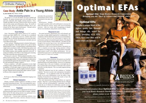
History and Presenting Symptoms A 28-year-old male presents with recurring episodes of moderate low-back pain which always respond well to chiropractic adjustments, but eventually return. He recalls no specific back injuries, and cannot identify any triggering activities. On a Visual Analog Scale, he rates his low back pain currently at about 50mm. He has been able to avoid taking pain medication by getting regular chiropractic adjustments. Exam Findings Vitals. This active young man is 6 feet tall and weighs 176 lbs, resulting in a BMI of 24; his muscle definition indicates that he is not at risk of overweight. He doesn't smoke, and his blood pressure is 116/78 mmHg with a pulse rate of 68 bpm. These findings are within the healthy range. Posture and gait. Standing postural evaluation finds a lower right iliac crest and a low right greater trochanter. His knees are well aligned, but there is obvious medial bowing of the right Achilles tendon, and no medial arch on the right foot. When this is mentioned, he recalls a "bad sprain" injury to his right foot and ankle during high school football. He denies any persisting symptoms or current problems with the right foot or ankle. Chiropractic evaluation. All active spinal ranges of motion are full and pain free, except that "aching stiffness" restricts left lateral flexion by 10°. Active palpation identifies a motion limitation in the right SI joint, which is also tender to direct pressure. Lumbosacral joint motion is restricted in left lateral flexion, with the feeling of generalized paraspinal muscle tightness. Provocative orthopedic and neurological tests for nerve root impingement and disc involvement are all negative. Lower extremities. Closer examination reveals that he has no right medial arch when standing. His right calcaneus is noticeably everted when bearing weight. When seated and non-weightbearing, his right arch appears equal to the left, and manual testing finds no evidence of muscle weakness of the fibular or tibial muscles. The Navicular Drop test measures substantial asymmetry in excursion of the navicular bones when moving from sitting to standing (R = 3 mm of drop, L = 8 mm of drop from non-weight bearing to weight bearing). Palpation finds no significant tenderness in the right medial arch or plantar fascia. Imaging A lumbopelvic series (AP and lateral lumbopelvic views) reveals an obvious discrepancy in femur head heights, with a measured difference of 5 mm (right side lower). A moderate lumbar curvature (6°) is noted, convex to the right side, and both the sacral base and the iliac crest are lower on the right side. The sacral base angle and measured lumbar lordosis are within normal limits. Clinical Impression Asymmetrical pronation likely due to previous injury, with associated pelvic tilt and lumbar curvature resulting in chronic biomechanical stress and recurring subluxations in the lumbo-pelvic region. Treatment Plan Adjustments. Specific, corrective adjustments for the SI and lumbosacral joints were provided—with good response, as previously. Manipulation of the right foot, including the navicular, cuboid, and calcaneal bones, was also performed. Support. Custom-made, flexible stabilizing orthotics were supplied, with a pronation correction added to the right side. He had no problems in wearing the orthotics, finding them "very comfortable." Rehabilitation. He was shown a series of lumbopelvic mobility exercises, using elastic exercise tubing at home. He was encouraged to continue his twice-weekly workouts at the local gym. Response to Care The lumbopelvic and foot adjustments were well tolerated, and the orthotics made a noticeable improvement in his postural alignment, both at the feet and the lumbopelvic region. After 6 weeks of adjustments (12 visits) and daily home exercises, including wearing the orthotics, he was released to a self-directed maintenance program. Discussion Excessive pronation and biomechanical asymmetries in the foot and ankle are often locally asymptomatic. In this active patient, the constant weightbearing stress to his SI and lumbosacral joints resulted in recurring spinal symptoms. Preventing chronicity is a vital aspect of chiropractic, and correction of this patient's underlying pronation asymmetry was necessary. Having had no foot or ankle symptoms, he had not recognized that a previous lower-extremity sports injury could be a significant causative factor in his back problem. Fortunately, this was identified before any substantial degenerative changes developed. Dr. JohnJ. Danchik is the seventh inductee to the American Chiropractic Association Sports Hall of Fame. He is the current chairperson of the United States Olympic Committee s Chiropractic Selection Program and lectures extensively in the United States and abroad on current trends in sports chiropractic and rehabilitation. Dr. Dan- chik is an associate editor of the Journal of the Neuromusculoskeletal System. He can be reached by e-mail at docforjocs@aol. com. \