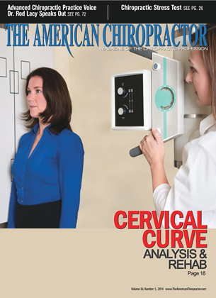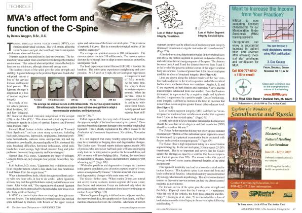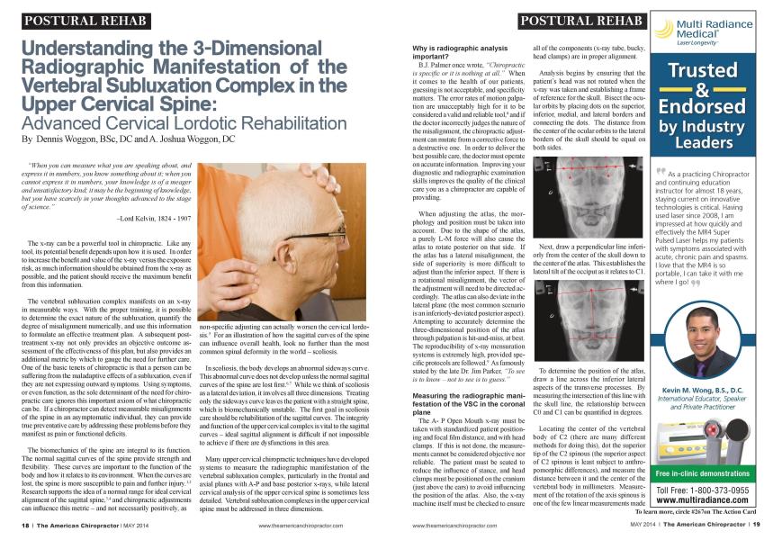
"When you can measure what you are speaking about, and express it in numbers, you know something about it; when you cannot express it in numbers, your knowledge is of a meager and unsatisfactory kind; it may be the beginning of know ledge, but you have scarcely in your thoughts advanced to the stage of science." -Lord Kelvin. 1824- 1907 The x-ray can be a powerful tool in chiropractic. Like any tool, its potential benefit depends upon how it is used. In order to increase the benefit and value of the x-ray versus the exposure risk, as much information should be obtained from the x-ray as possible, and the patient should receive the maximum benefit from this information. The vertebral subluxation complex manifests on an x-ray in measurable ways. With the proper training, it is possible to determine the exact nature of the subluxation. quantify the degree of misalignment numerically, and use this information to formulate an effective treatment plan. A subsequent post-treatment x-ray not only provides an objective outcome assessment of the effectiveness of this plan, but also provides an additional metric by which to gauge the need for further care. One of the basic tenets of chiropractic is that a person can be suffering from the maladaptivc effects of a subluxation. even if they arc not expressing outward symptoms. Using symptoms, or even function, as the sole determinant of the need for chiropractic care ignores this important axiom of what chiropractic can be. If a chiropractor can detect measurable misalignments of the spine in an asymptomatic individual, they can provide tme preventative care by addressing these problems before they manifest as pain or functional deficits. The biomcchanics of the spine are integral to its function. The normal sagittal curves of the spine provide strength and flexibility. These curves arc important to the function of the body and how it relates to its environment. When the curves are lost, the spine is more susceptible to pain and further injury.1-2 Research supports the idea of a normal range for ideal cervical alignment of the sagittal spine."'4 and chiropractic adjustments can influence this metric - and not necessarily positively, as non-specific adjusting can actually worsen the cervical lordo-sis.5 For an illustration of how the sagittal curves of the spine can influence overall health, look no further than the most common spinal deformity in the world - scoliosis. In scoliosis. the body develops an abnormal sideways curve. This abnoniial curve docs not develop unless the normal sagittal curves of the spine arc lost first.''" While we think of scoliosis as a lateral deviation, it involves all three dimensions. Treating only the sideways curve leaves the patient with a straight spine, which is biomcchanically unstable. The first goal in scoliosis care should be rehabilitation of the sagittal curves. The integrity and function of the upper ccnical complex is vital to the sagittal cun cs - ideal sagittal alignment is difficult if not impossible to achieve if there arc dysfunctions in this area. Mam upper ccnical chiropractic techniques have developed systems to measure the radiographic manifestation of the vertebral subluxation complex, particularly in the frontal and axial planes with A-P and base posterior x-rays, while lateral ccnical amilvsis of the upper ccnical spine is sometimes less detailed. Vertebral subluxation complexes in the upper cenical spine must be addressed in three dimensions. Why is radiographic analysis important? B.J. Palmer once wrote. "Chiropractic is specific or it is nothing at all. " When it comes to the health of our patients, guessing is not acceptable, and specificity matters. The error rates of motion palpation arc unacceptable high for it to be considered a valid and reliable tool." and if the doctor incorrectly judges the nature of the misalignment, the chiropractic adjustment can mutate from a corrective force to a destructive one. In order to deliver the best possible care, the doctor must operate on accurate information. Improving your diagnostic and radiographic examination skills improves the quality of the clinical care you as a chiropractor arc capable of providing. When adjusting the atlas, the morphology and position must be taken into account. Due to the shape of the atlas, a purely L-M force will also cause the atlas to rotate posterior on that side. If the atlas has a lateral misalignment, the side of superiority is more difficult to adjust than the inferior aspect. If there is a rotational misalignment, the vector of the adjustment will need to be directed accordingly. The atlas can also deviate in the lateral plane (the most common scenario is an infcriorly-dcviatcd posterior aspect). Attempting to accurately determine the three-dimensional position of the atlas through palpation is hit-and-miss, at best. The reproducibility of x-ray mensuration systems is extremely high, provided specific protocols are followed.1' As famously stated by the late Dr. Jim Parker. "To see is to know - not to see is to guess. " Measuring the radiographic manifestation of the VSC in the coronal plane The A- P Open Mouth x-ray must be taken with standardized patient positioning and focal film distance, and with head clamps. If this is not done, the measurements cannot be considered objective nor reliable. The patient must be seated to reduce the influence of stance, and head clamps must be positioned on the cranium (just above the cars) to avoid influencing the position of the atlas. Also, the x-ray machine itself must be checked to ensure all of the components (x-ray tube, bucky. head clamps) are in proper alignment. Analysis begins by ensuring that the patient's head was not rotated when the x-ray was taken and establishing a frame of reference for the skull. Bisect the ocular orbits by placing dots on the superior, inferior, medial, and lateral borders and connecting the dots. The distance from the center of the ocular orbits to the lateral borders of the skull should be equal on both sides. Next, draw a perpendicular line inferi-orly from the center of the skull down to the center of the atlas. This establishes the lateral tilt of the occiput as it relates to C1. To determine the position of the atlas, draw a line across the inferior lateral aspects of the transverse processes. By measuring the intersection of this line with the skull line, the relationship between CO and Cl can be quantified in degrees. Locating the center of the vertebral body of C2 (there arc many different methods for doing this), dot the superior tip of the C2 spinous (the superior aspect of C2 spinous is least subject to anthropomorphic differences), and measure the distance between it and the center of the vertebral body in millimeters. Measurement of the rotation of the axis spinous is one of the few linear measurements made with a ruler rather than a protractor. It will not be valid if the FFD on the x-rays being compared is different. The next step is to analyze the position of the atlas as it pertains to the cervical spine. Draw a line from the center of C2 to the center of C5; extend this line a little farther up to intersect with the line drawn across the lateral, inferior aspects of the atlas. Measuring the intersection made by these two lines on the side of the acute angle provides the radiographic manifestation of the lateralitv of the atlas compared to the cervical spine. Measuring the radiographic manifestation of the VSC in the axial plane Axial measurement of the radiographic manifestation of the VSC requires obtaining a Base Posterior x-ray (which is similar to a vertex or Water's view, but in a BP x-ray, the bucky is not tilted). This x-ray must be taken with standardized patient positioning and focal film distance, and with head clamps. This ensures that measurements arc accurate and unifonn across prc- and post- treatment x-rays. Bisecting the glabclla and the center mass of the dens provides a frame of reference for the cranium. Draw ing a line across the posterior aspect of the lateral masses of the atlas (or through the center of the transverse foramina, if the lateral masses are difficult to visuali/c) then provides an objective measurement of the orientation of the atlas. The anteriority or posteriority of the atlas is referenced on the side of atlas latcrality and expressed in degrees. Measuring the radiographic manifestation of the VSC in the lateral plane The measurement of the upper cervical spine in the lateral plane is absent from mam upper cervical techniques. This is an important part of understanding the three-dimensional nature of the VSC. This is the analysis of the lateral cervical neutral x-ray for the CLEAR protocol. The red lines measure loss of curve (LOC). quantified as a percentage. The blue line measures forward head posture (FHP). measured in inches and expressed as an increase in the apparent weight of the head (AHW). The green lines measure post-articular dysfunction (PAD), and loss of motion segment integrity (LMSI) is evaluated as well for signs of dysfunction in the ALL or PLL. The yellow lines measure the upper cervical complex in the lateral plane. The C2 vertebral body base line has a specific relationship with the cervical lor-dosis. In a normal sagittal curve, the C2 vertebral body base line intersects a pure horizontal at ten degrees inferior (-10 ). In a patient with a cervical kyphosis, the C2 vertebral body base line will intersect the horizontal plane line superiorly (in this example. +24 . which is 34 degrees of misalignment). Once all three dimensions of the upper cervical complex have been analyzed, it is possible to quantify the total misalignment present in all three planes. Consider the following example: The occiput is tilted 6 degrees. The atlas is misaligned by 2 degrees in the coronal plane. 6 degrees axially. and 24 degrees laterally. C2 itself (sen ing as the foundation of the atlas) is misaligned by 38 degrees. This gives a total of 82 degrees of 3-dimcnsional upper cervical misalignment; this is a very significant amount of misalignment, all occurring in one concentrated and vital area! Until this can be reduced, it is highly unlikely that the alignment of the spine below can be definitively addressed. Gravity works from the top-down, and the righting reflex of the body will always align the eyes to a pure horizontal: any misalignments present in the atlas will cause this system of orientation to be thrown into chaos as the brain compensates to maintain an upright and level position. One example of the ramifications of lateral C2 subluxation in sagittal corrective care The position of the C2 vertebral body base line is not only a scgmcntal subluxation. but rather has a global effect due to the kinetic chain of the spine. This can be explained through physics and biomcchanics. The scllac turcica represents the center of mass of the skull. It is located anterior to the articulating surfaces of the occiput. atlas, and axis. The position of the skulls center of mass has a cantilever effect upon the cervical spine. When the center of mass is shifted forward, the weight and gravitational forces transfer through the C2 vertebral body and the cervical spine goes into a kyphosis. By using the C2 spinous as a lever, the center of mass of the skull can be translated posteriorly, reducing the cantilever effect and aiding in the restoration of the cervical lordosis. This adjustment can be performed with a mechanical adjusting instrument using a single-prong stylus. The patient Ilexes their head while seated, as the doctor places the stylus on the superior, posterior aspect of the C2 spinous. Adjustments are delivered at a frequency of 6 Hz as the doctor brings the patient's head into a slight extension. The line of drive is predominantly P-A (as the morphology of the axis ensures that, as the patient's head goes into extension, the forces will transmit S-I). Why is mechanical adjusting instrument adjusting important? While there arc a few manual and drop-assisted adjustments that arc sometimes used to influence the alignment of the upper cervical complex, in my clinical experience, none of these techniques can match the specificity and accuracy of the stylus of a mechanical adjusting instrument. Almost all of the chiropractic techniques which utilize prc- and post- x-rays as an outcome assessment utilize some form of mechanical adjusting instrument (Grostic is one of the few exceptions). While spring-loaded adjusting instruments have their applications and benefits, two advantages of mechanical instruments arc the rapidity and speed with which the adjustment can be delivered, as well as the consistency of the force. A spring-loaded instrument loses force as it travels, much like a bow and arrow. Some mechanical adjusting instruments can also deliver 6 pulses per second, which is the equivalent of one adjustment every 167 milliseconds. The nervous system reacts to stimuli at approximately 333 milliseconds: this means that the corrective force can be delivered faster than the body can react to it. Research has found that at this frequency (6 Hz), the amount of vertebral motion that can be achieved is greatly enhanced.10 Mam factors must be addressed to successfully rehabilitate a loss of the cervical lordosis. Sometimes missing even just one of these factors can be enough to render the treatment ineffective. The maximum potential of the chiropractic adjustments is not limited by the clinical skills of the doctor, but by the gaps in their diagnostic procedures. A doctor cannot treat the problems he or she docs not identify. The upper cervical complex must be anah zed and corrected in all three dimensions, and the soft tissue and neurological aspects must be considered and treated as well. As experts in spinal pathophysiology. Doctors of Chiropractic should avail themselves of all the tools that may help health and function of their patients. When utilized responsibly and ethically, x-ray analysis can improve the specificity and accuracy of the chiropractic adjustment, and thus your chances of achieving a successful treatment outcome in your patients. References Stcmper BD. Yoganandan N. Pintar FA: Effects of abnormal posture on capsular ligament elongations in a computational model subjected to whiplash loading. J Biomcch 2005 Jim: 38(6) 1313-23. McAvincy J ct al: Determining the Relationship Between Cervical Lordosis and Neck Complaints. JMPT March-April 2005: Vol 28 No. 3 Harrison DD. Troyanovich SJ. Harrison DE. Janik TJ. Murphy DJ: A normal sagittal spinal configuration: a desirable clinical outcome. JMPT 1996 Jul-Aug:19(6):398-405 4. Harrison DD 1. Janik TJ. Troyanovich SJ. Holland B: Comparisons of lordotic cervical spine curvatures to a theoretical ideal model of the static sagittal cervical spine. Spine (Phila Pa 1976). 1996 Mar 15:21(6):667-75. 5. Troyanovich SJ. Harrison DD. Harrison DE: A review of the valid- ity, reliability, and clinical effectiveness of chiropractic methods employed to restore or rehabilitate cervical lordosis. Chiropr Technique. 1998:10(1): 1-7. 6. Roussouly PI. Labellc H. Rouissi J. Bodin A: Prc- and post operative sagittal balance in idiopathic scoliosis: a comparison over the ages of two cohorts of 132 adolescents and 52 adults. Eur Spine J. 2013 Mar.22 Suppl 2:S2O3-15. doi: 10.1007/s00586- 012-2571-x. Epub 2012 Nov 28. 7. Lawlon JO. Dickson RA: The experimental basis of idiopathic scoliosis. Clin Orthop Rclat Res. 1986 Scp:(210):9-17. 8. Stochkcndahl MJ. Christcnscn HW. Harlvigscn J. Vach W. Haas M. Hcslback L. Adams A. Bronfort G: Manual examination of the spine: a systematic critical literature review of rcproducibilily. JMPT 2006 Jul-Aug:29(6):475-85. 485c 1-10 9. Harrison DE 1. Harrison DD. Colloca CJ. Bctz J. Janik TJ. Holland B: Repeatability over time of posture, radiograph positioning, and radiograph line drawing: an analysis of six control groups. JMPT 2003 Feb:26(2):87-98. 10. Keller TS1. Colloca CJ. Moore RJ. Gun/burg R. Harrison DE: Increased multiaxial lumbar motion responses during multiple-impulse mechanical force manually assisted spinal manipulation. Chiropr Osteopat. 2006 Apr 6; 14:6. Dr. Dennis Woggon is the founder of the CLEAR Scoliosis Institute and the St. Cloud Chiropractic Clinic. lie graduated from Palmer College oft 'hiropraciic in 1974. lie is an international instructor for CI.t'AR Scoliosis Institute. He can be contacted at dnvoggonfclclear-institute.org. Dr. A. Joshua II oggon, a 2010 Cjrachiale ofl'arker ( allege, serves as the Director of Research for the CLEAR Scoliosis Institute, a Xon-I'rofit Organization dedicated lo aih'ancing chiropractic scoliosis correction (nww.clear-inslilule.org). lie can be contacted at jwoggon'&clear-institiite.org.