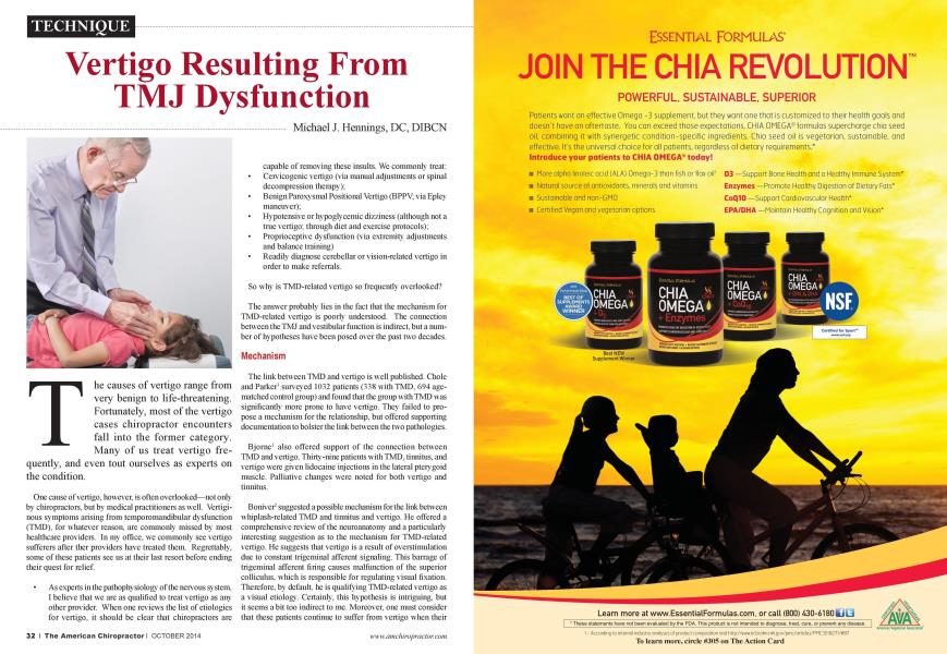
T he causes of vertigo range from very benign to life-threatening. Fortunately, most of the vertigo cases chiropractor encounters fall into the former category. Many of us treat vertigo frequently, and even tout ourselves as experts on the condition. One cause of vertigo, however, is often overlooked—not only by chiropractors, but by medical practitioners as well. Vertiginous symptoms arising from temporomandibular dysfunction (TMD). for whatever reason, arc commonly missed by most healthcare providers. In my office, we commonly sec vertigo sufferers after ther providers have treated them. Regrettably, some of these patients sec us at their last resort before ending their quest for relief. As experts in the pathophysiology of the nervous system. I believe that we arc as qualified to treat vertigo as any other provider. When one reviews the list of etiologies for vertigo, it should be clear that chiropractors arc capable of removing these insults. We commonly treat: Ccn icogenic vertigo (via manual adjustments or spinal decompression therapy): Benign Paroxysmal Positional Vertigo (BPPV; via Epley maneuver): Hypotcnsivc or hypoglyccmic dizziness (although not a tnic vertigo: through diet and exercise protocols): Proprioccptive dysfunction (via extremity adjustments and balance training) Readily diagnose ccrcbcllar or vision-related vertigo in order to make referrals. So why is TMD-related vertigo so frequently overlooked? The answer probably lies in the fact that the mechanism for TMD-rclatcd vertigo is poorly understood. The connection between the TMJ and vcstibular function is indirect, but a number of hypotheses have been posed over the past two decades. Mechanism The link between TMD and vertigo is well published. Chole and Parker5 surveyed 1032 patients (338 with TMD. 694 age-matched control group) and found that the group with TMD was significantly more prone to have vertigo. They failed to propose a mechanism for the relationship, but offered supporting documentation to bolster the link between the two pathologies. Bjorne1 also offered support of the connection between TMD and vertigo. Thirty-nine patients with TMD. tinnitus, and vertigo were given lidocainc injections in the lateral ptcrygoid muscle. Palliative changes were noted for both vertigo and tinnitus. Bonivcr2 suggested a possible mechanism for the link between whiplash-related TMD and tinnitus and vertigo. He offered a comprehensive review of the ncuroanatomy and a particularly interesting suggestion as to the mechanism for TMD-rclatcd vertigo. He suggests that vertigo is a result of ovcrstimulation due to constant trigeminal afferent signaling. This barrage of trigeminal afferent firing causes malfunction of the superior colliculus. which is responsible for regulating visual fixation. Therefore, by default, he is qualifying TMD-relatcd vertigo as a visual etiology. Certainly, this hypothesis is intriguing, but it seems a bit too indirect to me. Moreover, one must consider that these patients continue to suffer from vertigo when their eves are closed, a not typically offered by visual-related vertigo sufferers. More likely, one must consider the changes to the pharyngo-tvmpanic tube as a result of TMD. The TMJ is innervated by the trigcminal nerve4, which also innervates two pharyngotym-panic muscles—tensor tympani and tensor vcli palatini. Likely, aberrant function of the trigcminal nerve resulting from TMD leads to improper tone in these muscles leading to increased intra-auricular pressure. Correlation between increased intra-auricular pressure and vertigo has been made by Montandon. ct al.'1 by documenting relief from vertigo by use of transtym-panic ventilation tubes. Although Montandon docs not propose a mechanism for increased pressure in causing vertigo, one could consider that this pressure disnipts proper endolymphatic flow. This hypothesis also parallels the purpose of performing caloric reflex testing in the external auditor)' meatus~thc hot or cold water creates convection of the cndolj mphatic fluid leads to vertigo. The most likely mechanism for TMD-rclated vertigo, in my opinion, is related to the niechanoreceptors within the TMJ. The high concentration of niechanoreceptors in the TMJ makes the TMD patient vulnerable to proprioceptivc problems. Kokai. ct al.5 supports this hypothesis in an animal study in which rats" mandibles were passively deviated laterally. Elcctrophsiologic changes (lower firing thresholds) were recorded from the joints" niechanoreceptors versus those in the control groups. Certainly, vertigo is a likely sequela from aberrant motion of the TMJ. particularly when one considers the nearly constant motion of the joints when talking, chewing or breathing. Furthermore, the trigcminal nerve synapses at the spinal trigcminal nucleus, which communicates extensively with the cerebellum via trigcininoccrcbcllar connections through the superior ccr-ebcllar peduncle4. The facial nerve may also play a part in this scenario, due to its connection with the spinal trigcminal tract. The facial nerve exits the cranium through the stvlomastoid foramen7. Theoretically, the juxtaposition of this foramen to the TMJ makes the facial nerve susceptible to local inflammatory insult from the joint and surrounding soft tissues. Exam There are countless tests that can be performed to pinpoint causes for vertigo/dizziness. However, my routine exam consists of a few handpickcd orthopedic and neurological tests which arc typically enough to find the etiology for vertigo. Typically. I start with palpation of the cervical spine. Ccrvicogcnic vertigo may be elicited by this maneuver alone, or may require range of motion of the joints to cause symptoms. If no symptoms arc reproduced. I then perform Dix-Hallpikc maneuver. The patient is seated with head rotated toward the examiner and eyes open. He/she is asked to lie back so that the head is extended off the end of the table. If nystagmus is elicited, this position can transition right into Eplcy "s maneuver (description is outside the scope of this article). Another clever test to identify' vertigo etiology is the "Swivel Chair Test" (hat tip. Joseph S. Fcrczy. DC). The patient is seated on a swivel (examination) chair and passively rotated 180 degrees repeatedly. The intent is to test visual or vcstibular-evoked vertigo. If no nystagmus is elicited, the procedure is repeated with the patient's head held stationary while the body swivels. This portion of the test excludes any vcstibular or visual etiologies, and reproduction of vertigo is confirmatory of cervicogenic vertigo. As an aside, an otolaryngologist taught me one question that should be asked of even vertigo patient. He said that he always asks if the patient gets up to use the bathroom in the middle of the night, and if so. do they turn on the lights? His logic is that, since most people use the bathroom at night and almost never turn on the lights, if they still have vertigo it is not likely of visual origin since they are not dependent on the visual system at that time. Lastly, if no other tests arc provocative for tlicir vertigo, palpate the TMJ bilaterally and try some excursion of tlie mandible. If the patient is supine and otherwise stationary, any vertigo provoked with this palpation is confirmatory forTMD etiology. Manual reduction of this subluxation should immediately abate the patient's symptoms. Soft tissue work may be necessary. My personal favorite approach is a simple axial traction of the mandible. Both thumbs are placed inside tlie patient's mouth resting on the bottom teeth and a gradual downward (caudal) pressure is applied. Patients arc tolerant of this maneuver and it rarely provokes complaint. Because of tlie mechanical etiology of TMD-rclatcd vertigo, clearly cliiropractors sliould serve as the first line of defense for this type of symptom. Manual reduction of temporomandibular subluxation is common practice in cliiropractic offices, altliougli tlie intention is usually palliative treatment for pain rather than vertigo. With the help of the above-listed differential tests, the chiropractor that diagnoses and treats TMD-rclated vertigo is sure to be hcro-for-a-day—hands down tlic best way to grow a successful practice! Dr. S lichael Hennings is a Chiropractic Neurologist board certified by the International Board of Chiropractic Xeurology. He received his bachelor s degree in biology from the University of South Florida followed by a Doctorate of Chiropractic Medicine from Palmer C "allege o/Chiropractic Florida. He is currently in private practice in Palm City, FL. He can be reached at 772-220-5880 or mhennings l@hotmail. com. References: /. /. Bjorne. A. (2007). Assessment of temporomandibular disorder and cen'ical spine disorders in tinnitus patients. Progress in Brain Research: \bl 166, pp 215-219. Boniver, R. (2002). Temporomandibular joint dysfunction in whiplash injuries: Association with tinnitus and vertigo. In ternational Tinnitus Journal. I'olS, Xo. 2, pp. 129-131. Chole, R. A., and Parker, \V. S. Richard A. (1992). Arch Oto- laryngol Head Neck Surg. 1992; U8(8):817-821. Haines, D. E. (2002). Fundamental Xeuroscience: Second Edition. Philadelphia, PA: Churchill Livingstone. Kokai. S., Yabushita. T. V.eredo, .1. I... Toda K., andSoma. K. (2007). Functional Changes of the Temporomandibular Joint Mechanoreceptors Induced by a Lateral Mandibular Shift in Rats. The Angle Orthodontist: May 2007, Vol. 77. Xo. 3. pp. 436-441. Monlandon, I'.. Cruil/emin, P.. andIldusler. R. (19HH). Preven tion of I erligo in Meniere s Syndrome by Means ofTranstym- panic Ventilation Tubes. ORL. Vol. 50, Xo. 6. Xetter, F. H. (2006). Atlas of Human Anatomy: Fourth Edition. Philadelphia, PA: Sounders Elsevier.