The Theory of Spinal Compression Syndrome (SCS) as it relates to Subluxation: Mechanism of Injury or Diagnosis
PERSPECTIVE
David B. Bass
DC, LAC, AP, DOM
With all of the ongoing changes in diagnostic coding, we are faced many times with using a code that “almost fits” the patient’s condition. In this article, I’d like to discuss spinal compression syndrome as a diagnosis, not simply a mechanism of injury.
What is spinal compression syndrome (SCS)? I believe we can all agree that aggressive vertical compressions to the spine can and do create serious spinal injuries ranging from compression fractures to wedge compression subluxations. However, to what extent does this mechanism of injury become a diagnosis?
For example, if you can diagnose facet imbrication (overclosure of the facets), we look for a suitable code, but what caused the facets to imbricate? Could SCS be the etiology? How about a hyperextension injury (which is also a compression)? Let me develop the theory.
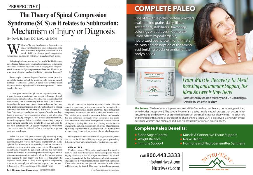
As the spine moves through normal day-to-day activities, it goes through a continuous and repetitive barrage of axial compressing and rebounding. A healthy disc can provide all of the necessary spinal rebounding that we need. This rebounding enables the spine to recover to its vertical neutral, but over time, continuous compression and rebounding begin to fatigue the bonds that maintain the integrity of the disc’s annular fibers. Once this process begins, the layers of annular cartilage begin to separate. This weakens disc integrity and allows the process of bulging to begin. As this process gains momentum, the nucleus pulposus follows behind the annular bulge, preparing to herniate once the outer annular fibers tear and open. In this case, a diagnosis of SCS could be appropriate because the compression must be relieved before a lasting disc improvement can be achieved.
When you observe a spine with osteophytes running along multiple vertebrae segments, the typical diagnosis would be osteophytosis, but could it be evidence of chronic SCS? In my opinion, the osteophytes are a secondary condition resultant of multiple repetitive vertical axial compressions. This repetitive microtrauma weakens the periosteal cartilage that surrounds the vertebral bodies. It causes the periosteal cartilage to flap off at the compression points, typically where the bodies meet the disc. Because the body doesn’t like these loose flaps, the body begins to calcify them. As long as the repetitive compressing remains, the osteophytes will continue to grow. Here we have a diagnosis of SCS complicated with osteophytosis.
Not all compression injuries are vertical axial. Flexionextension injuries are just as compressive. In the typical forward-impact auto-related trauma, an extreme flexion movement compresses the anterior vertebral bodies and anterior discs. The reactive hyperextension movement injures the posterior disc and imbricates the facets. When the anterior and posterior vertebrae and disc remain compressed, we turn vertebral gliding into grinding. Over time, the grinding reveals itself as osteoarthritis and disc degeneration. This type of compression injury may respond better if decompression was administered to remove any compressions between the vertebral segments.
Although there is a flexion-extension diagnostic code, in this case, a code for SCS would be just as appropriate, particularly if decompression was a component of the therapy program.
MRIs and SCS
We always review MRIs before confirming disc involvement. As such, many times we see normal disc spacing without bulging. However, in the T-2 images, the absence of a whitish color in the center of the disc indicates a dehydration process. The disc needs movement for imbibition and hydration to occur. When a disc becomes compressed, the vertebral units above and below may be fixated. This stops the imbibition process,
resulting in disc desiccation with annular fiber weakening. In this situation, I believe that chronic compression is both the cause and diagnosis that predisposes the disc to future problems. Decompression should be used to release the fixated vertebral segments and to create a negative disc vacuum for renewed disc hydration.
Subluxation and Spinal Compression Syndrome
Is it possible that subluxations are compensatory distortions of spinal compression syndrome? As the spine begins to compress, discs become squeezed and pressured. Simultaneously, vertebral segments will react by moving in a variety of ways to avoid compression. These include wedge compression subluxations as well as flexion-extension and rotational malpositions. Once repetitive compressive forces overwhelm the discs and facets, the ability of the column to rebound becomes difficult. Vertebrae subluxate to “get out of the way” of any compressive force. If this is the case, the subluxation listing becomes chr onic until the compression is removed, and the subluxation is secondary to SCS.
To conclude, if the theory of SCS is valid, compressions must be released to maximize improvement and rehab.
Dr David Bass is both a Chiropractic andAcupunclure Physician. He is currently the owner and director of the Neck and Back Pain Institute of Coral Springs and is also President of Big Rehab Corp., the manufacturer ofthe FDA-Cleared Antalgic-Trak ROM Spinal Decompression System. He can be reached at: ContacCcfAntalgic-Trak.com