Body Sway, Proprioception, and Spinal Curves
SCOLIOSIS BODY SWAY
Richard M. Opper
DC
Body sway” is defined as a movement of the upper body as if it were an upside-down pendulum. It is present primarily when one is standing in an upright and static posture and is noticeable when one places the feet together and closes the eyes. The swaying motion becomes more perceptible in that position. The degree of sway can be influenced by several factors: upper motor lesions, muscle pathology or injury, pathology or injury to spino-sensory or motor tracts, labyrinthine or ocular balance dysfunction, changes in skeletal morphology, joint injury or disease, dysfunctional pedal mechanics, and environmental stresses.
The degree of sway is quantifiable by using instrumentation and can be useful information in diagnosing neuromuscular disorders. There are normal variants in the degree of sway. For example, it is found to increase in people over 60 years of age. When the degree of sway becomes moderately or severely evident, the terms “disequilibrium” or “standing unbalance” are used to describe the condition.
Body sway is the direct consequence of autonomic balance mechanisms. These mechanisms are constantly activated when we are upright, standing still, or in motion. The human body is structurally unstable and requires powerful mechanisms to maintain upright balance. The most powerful of these mechanisms is located in the viscoelastic tissues of the spinal column and the deep muscles and fascia of the back. A continual contraction/ relaxation of these deep muscles in the back attempts to maintain the body ’ s center of mass on or near the vertical gravity line. When the body is leaning too far forward, the deep muscles of the back will contract and extend the spine in order to shift the center of mass backward. If the body is leaning too far to the right or left, the deep spinal muscles contract and exert a lateral force on the spinal column to accomplish the same thing. This activity is the powerful neuromuscular spinal reflex commonly called “the righting reflex.”
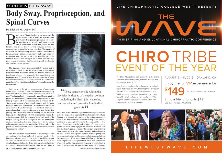
The most abundant concentration of proprioceptive sensors outside of the head and neck is in the vicinity of the lower thoracic and upper lumbar spine in the kidney region (1). These sensors reside within the viscoelastic tissues of the spinal column, including the discs, joint capsules, and anterior and posterior longitudinal ligaments. They are also found in
"These sensors reside within the viscoelastic tissues of the spinal column, including the discs, joint capsules, and anterior and posterior longitudinal ligaments."
abundance in this particular region in the deep spinal muscles and in the fascia. They are primarily mechanoreceptors whose function is to transmit infonnation to the brain regarding the status of the body’s balance. More specifically, these sensors, also known as graviceptors(1), monitor the position of the body’s center of mass relative to the body’s center of pressure on the ground. So when one is upright and balanced, a vertical line from the body’s center of mass, which is just anterior to the sacral plateau, will reach the ground at a point midway between the feet. The area on the ground bundied by an imaginary line encompassing the feet and the space in between is called the “base of support.” When one is about to fall forward, the body’s center of pressure on the ground will be in front of the base of support, and the neuromuscular response, prompted by the sensors, will attempt to change the body’s posture in order to
reposition the body’s center of mass back toward the center of the base of support. It accomplishes this by triggering contraction of the deep back muscles, which extend the spine and rotate the pelvis backward, thus influencing the body’s center of mass to also move backward (2). There are circumstances in which the proprioceptors are stimulated continuously with the consequence of prolonged or chronic deep back muscle contraction, which may be in extension, lateral flexion, or rotation, or any combination of those (2).
Studies performed on children with untreated adolescent idiopathic scoliosis revealed the prevalence and increase of body sway (disequilibrium), which coincided with the onset of scoliosis and ceased abruptly when the lateral curves were fully formed(3). The spinal curves in the sagittal plane were observed to have changed dramatically along with the lateral curving in the fr ontal plane. Researchers who examined the spines in vitro of people with untreated and fully formed idiopathic scoliosis noticed that the lateral or sagittal view of the spine and pelvis suggested that the structure had been subjected to a strong and continual muscular contraction that had extended, rotated and laterally flexed the spine but appeared to be flat in that plane (sagittal)(4,5).
The question then became: What is the impetus for a straight spine in the fr ontal plane to become crooked while straightening the spine in the sagittal plane? None of the 5,201 studies on adolescent idiopathic scoliosis have produced an answer to that
question. What might the provocative stimulus be that prompts a chronic muscular contraction to dramatically change the shape of the spine in all three dimensions? Does the onset of a lateral scoliotic curve initiate disequilibrium or does disequilibrium prompt the lateral curve? Studies have ruled out genetic mutation and links, CNS lesions, muscle pathology, spinal nerve lesions, and metabolic or endocrine pathology. Might it be physiologic? This will be explored in a future article.
References
1. Karakaya MG, Karakaya 1C, Karakaya DG; “Proprioception: The Forgotten Sixth Sence ”ch : spine and proprioception1 , OM1CS Group eBooks; May 2015
2. Opper RM, 20181 “Asolescent Idiopathic Scoliosis is an Etiotropic Remodeling of The Spine in Response to Disequilibrium; an Hypothesis ”, unpublished.
3. Yamada K, Yamamoto H, Nagakawa Y,et al. ; Etiology of idiopathic scoliosis; Clin ortho and related research; no. 184, 1984, Tokushima U, School ofnedicine
4. Roaf; The basic anatomy of scoliosis. J Borne Joint Surg. 48B: 786, 1966
5. Lawton J O, Dickson R A : The experimental basis of idiopathic scoliosis;jclin ortho and related research, no 210, ( Sept 1986
1969 graduate of The Columbia Institute of Chiropractic, Partner in Palmer Chiropractic PC, Palmer, Mass. Authored: "Paradoxical Motion of The Cervical Spine...." JACA, Jan, 2000; Article under review: "Adolescent Idiopathic Scoliosis is an Etiotropic Remodeling of The Spine in Response to Disequilibrium" www. drrichopperdc@gmail. com