The Reproductive System
ORGAN OF THE MONTH
Howard F. Loomis
Jr., DC
Introduction
This series of articles deals with the common symptoms and underlying problems caused by "stress” and the importance of understanding its many ramifications. Simply stated, stress (sympathetic stimulation) occurs any time the body has insufficient energy to meet the three essential demands placed on it, namely:
• Energy to oppose gravity (structure)
• Energy to maintain homeostasis in the extracellular fluids (visceral)
• Energy to maintain emotional and cognitive stability (brain)
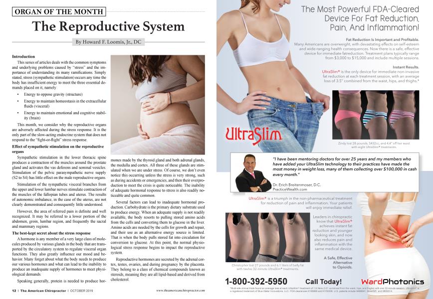
This month, we consider why the reproductive organs are adversely affected during the stress response. It is the only part of the slow-acting endocrine system that does not respond to the "fight-or-flight” stress response.
Effect of sympathetic stimulation on the reproductive organs
Sympathetic stimulation in the lower thoracic spine produces a contraction of the muscles around the prostate gland and activates the vas deferens and seminal vesicles. Stimulation of the pelvic parasympathetic nerve supply (S2 to S4) has little effect on the male reproductive organs.
Stimulation of the sympathetic visceral branches from the upper and lower lumbar nerves stimulate contraction of the muscles of the fallopian tubes and uterus. The results of autonomic imbalance, in the case of the uterus, are not clearly demonstrated and consequently little understood.
However, the area of referred pain is definite and well recognized. It may be referred to a lower portion of the abdomen, groin, lumbar region, and frequently the sacral and mammary regions.
The best-kept secret about the stress response
A hormone is any member of a very large class of molecules produced by various glands in the body that are transported by the circulatory system to regulate visceral organ functions. They also greatly influence our mood and behavior. Many forget about what the body needs to produce our various hormones and what can lead to the inability to produce an inadequate supply of hormones to meet physiological demands.
Speaking generally, protein is needed to produce hormones made by the thyroid gland and both adrenal glands, the medulla and cortex. All three of these glands are stimulated when we are under stress. Of course, we don't even notice this occurring unless the stress is very strong, such as during accidents or emergencies, and then their overproduction to meet the crisis is quite noticeable. The inability of adequate hormonal response to stress is also readily noticeable and quite common.
Several factors can lead to inadequate hormonal production. Carbohydrate is the primary dietary substrate used to produce energy. When an adequate supply is not readily available, the body resorts to pulling stored amino acids from the cells and converting them to glucose in the liver. Amino acids are needed by the cells for growth and repair, and their use as an alternative energy source is limited. That is when the body pulls stored fat into circulation for conversion to glucose. At this point, the normal physiological stress response begins to impact the reproductive system.
Reproductive hormones are secreted by the adrenal cortex, testes, ovaries, and during pregnancy by the placenta. They belong to a class of chemical compounds known as steroids, meaning they are all lipid-based and derived from cholesterol.
When we are under chronic or sustained stress, the body begins to use fats for energy, leaving less for sex hormone production. Slowly the ability to supply adequate lipids for production of reproductive hormones becomes a factor in the patient's symptoms as well as mood and behavior.
Obviously, the inability to adequately digest fats enters the symptomatic picture, such as when we have biliary problems. This happens regardless of whether we still have a
gallbladder and even occurs when we are on a ketogenic diet to lose weight. Our reserve of stored fat is being used for energy (instead of carbohydrate) and not as readily available for hormone production. This first becomes noticeable with the appearance of dry skin and excessive hair in the hairbrush.
Nutritional requirements for reproductive development
Lipids are precursors of prostaglandins and are used to stimulate
the production of follicle-stimulating hormones (FSH), luteinizing hormone (LH) or interstitial cell-stimulating hormone in males (ICSH) and lactogenic (LTH) hormone (prolactin) in women.
Embryology of the reproductive organs
The reproductive organs are developed from the mesoderm (as is the musculoskeletal system) and originate as a series of short outgrowths from the fifth cervical segment to the third thoracic segment. Each outgrowth segment then grows to the posterior and inferior, and they fuse together to form the Wolffian duct in the male or the Mullerian duct in the female. This duct continues to grow to the inferior until it opens into the front portion of the cloaca (the membrane that becomes the pelvic floor).
Sexual detennination depends upon the sex chromosomes X and Y and involves the specification of the gonads as either testes or ovaries. If the embryo is XY, the SRY gene (sex-determining) will be present. The protein produced by SRY activates a gene network that directs the gonads to develop as testes. In the absence of a Y chromosome and SRY, the gonads develop as ovaries.
The permanent organs of the adult are preceded by a set of structures that are purely embryonic and disappear almost entirely before the end of fetal life (except for the ducts leading to the exterior).
Male Development
Once the testes begin to develop, the two support cells in the testes differentiate and begin to generate important regulatory molecules that direct sexual differentiation.
• The Leydig cells produce testosterone, which promotes development of the Wolffian ducts. The Wolffian ducts then differentiate to form the epi-
didymis, vas deferens, seminal vesicles, and ejaculatory ducts.
• The Sertoli cells produce the Mullerian inhibiting substance (MIS), a peptide hormone that causes the Mullerian ducts to regress.
In the fetus, chorionic gonadotrophin (identical to LHICSH) is responsible for formation of male sexual organs.
FSH stimulates spermatogenesis in males and LH-ICSH from the pituitary stimulates the production of testosterone, which is needed for full maturity of the spermatozoa.
Female Development
Female development proceeds when there is an absence of the SRY gene. No testosterone or MIS is made. The Wolffian ducts regress, and the Mullerian ducts persist, developing into the fallopian tubes, uterus, and upper part of the vagina.
The female sexual cycle is completely dependent on gonadotrophic hormones from the anterior pituitary:
• Follicle-stimulating hormone (FSH) is required for the development and growth of the ovaries.
• Luteinizing hormone (LH) works in concert with FSH for follicular growth. Ovulation will not proceed without it, and it is responsible for maintaining secretions of the corpus luteum, namely estrogen and progesterone.
• Prolactin stimulates development of the breast and milk production after delivery.
Conclusion
When under stress (energy deficiency), the body resorts to using stored lipids as a replacement energy source. It pulls stored lipids into circulation (triglycerides), which reduces the level of lipids available for steroidal hormone production.
Next month, we look at visceral and structural stress and their effect on the kidneys.
Howard F. Loomis, Jr., DC, has an extensive background in enzymes and enzyme supplements. He is the founder and president of the Food Enzyme Institute™. His extensive knowledge of physiology, biochemistry, and enzymology has made him a sought-after speaker and a prolific writer. Dr. Loomis published ENZYMES: The Key to Health in 1999, as well as The Enzyme Advantage: For Healthcare Providers and People Who Care About Their Health, in 2015.
800-662-2630.