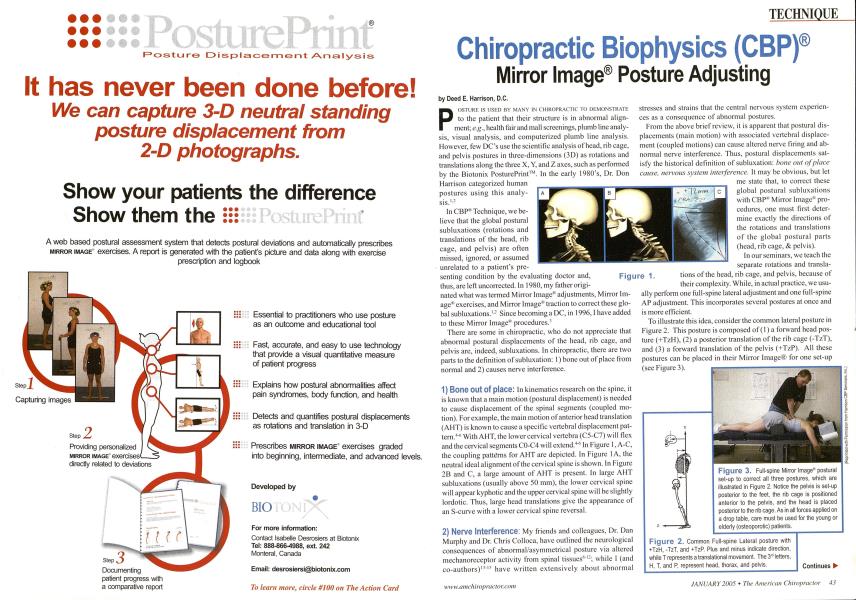
POSTURE IS USED BY MANY IN CHIROPRACTIC TO DEMONSTRATE to the patient that their structure is in abnormal alignment; e.g., health fair and mall screenings, plumb line analysis, visual analysis, and computerized plumb line analysis. However, few DCs use the scientific analysis of head, rib cage, and pelvis postures in three-dimensions (3D) as rotations and translations along the three X, Y, and Z axes, such as performed by the Biotonix PosturePrint™. In the early 1980's, Dr. Don Harrison categorized human postures using this analysis. '- In CBPR Technique, we believe that the global postural subluxations (rotations and translations of the head, rib cage, and pelvis) are often missed, ignored, or assumed unrelated to a patient's pre- senting condition by the evaluating doctor and, thus, are left uncorrected. In 1980, my father origi- nated what was termed Mirror Image" adjustments. Mirror Image" exercises, and Mirror Image" traction to correct these global subluxations.1 - Since becoming a DC, in 1996,1 have added to these Mirror Image* procedures.1 There are some in chiropractic, who do not appreciate that abnormal postural displacements of the head, rib cage, and pelvis are, indeed, subluxations. In chiropractic, there are two parts to the definition of subluxation: 1) bone out of place from normal and 2) causes nerve interference. 1) Bone Out Of place: In kinematics research on the spine, it is known that a main motion (postural displacement) is needed to cause displacement of the spinal segments (coupled motion). For example, the main motion of anterior head translation (AHT) is known to cause a specific vertebral displacement pattern.4'' With AHT, the lower cervical vertebra (C5-C7) will flex and the cervical segments C0-C4 will extend.46 In Figure 1,A-C, the coupling patterns for AHT are depicted. In Figure I A, the neutral ideal alignment of the cervical spine is shown. In Figure 2B and C, a large amount of AHT is present. In large AHT subluxations (usually above 50 mm), the lower cervical spine will appear kyphotic and the upper cervical spine will be slightly lordotic. Thus, large head translations give the appearance of an S-curve with a lower cervical spine reversal. 2) Nerve Interference: My friends and colleagues, Dr. Dan Murphy and Dr. Chris Colloca, have outlined the neurological consequences of abnormal/asymmetrical posture via altered mechanoreceptor activity from spinal tissues"12; while I (and co-authors)"15 have written extensively about abnormal stresses and strains that the central nervous system experiences as a consequence of abnormal postures. From the above brief review, it is apparent that postural displacements (main motion) with associated vertebral displacement (coupled motions) can cause altered nerve firing and abnormal nerve interference. Thus, postural displacements satisfy the historical definition of subluxation: bone out of place cause, nervous system interference. It may be obvious, but let me state that, to correct these global postural subluxations with CBP" Mirror Image11 procedures, one must first determine exactly the directions of the rotations and translations of the global postural parts (head, rib cage, & pelvis). In our seminars, we teach the separate rotations and transla- tions of the head, rib cage, and pelvis, because of their complexity. While, in actual practice, we usu- ally perform one full-spine lateral adjustment and one full-spine AP adjustment. This incorporates several postures at once and is more efficient. To illustrate this idea, consider the common lateral posture in Figure 2. This posture is composed of (1) a forward head posture (+TzH), (2) a posterior translation of the rib cage (-TzT), and (3) a forward translation of the pelvis (+TzP). All these postures can be placed in their Mirror Image® for one set-up (see Figure 3). Continues ► (Reprinted with Permission from Harrison CBP Seminars, Inc.) ► TECHNIQUE...From Previous Page Chiropractic Biophysics (CBP)® Mirror Image® Posture Adjusting (continued) Next, I wish to illustrate one common full-spine Mirror Image"1 set-up. Figure 4 illustrates a common AP posture, which is composed of a right low shoulder (right thoracic cage lateral flexion) and left head tilt (left lateral bending). Figure 5 depicts the CBP* Mirror Image" set-up/adjustment for this particular AP posture. In summary, Mirror Image"* postural set-ups/adjustments are unique in CBPrR> Technique. These methods were originated by Dr. Don Harrison in the early 1980's. These set-ups/adjustments are the exact opposite posture (or in difficult cases, these may be in a more stressed position) of the patient's initial presenting posture. While most doctors evaluate posture, they have not been taught the logical Mirror Image* methods that can result in routine postural correction. CBP" can make this claim of routine postural correction because we have investigated our methods with research designs. CBP" is the most published technique in the Index Medicus with over 80 published or in press research papers, of which six are Clinical Control Trials""21 and live are Case Studies (these are available online at www.idealspine.com). ► Go to www.TheAmericanChiropractor.com for References Deed E. Harrison, D.C., graduated from Ijfe-West Chiropractic College in IW6. He has authored nearly 71) peer reviewed research articles in journals such as: the JMPT. Spine, Clinical Biomechanics, etc. Dr. Harrison is a manuscript reviewer for the orthopedic journals Spine and Clinical Anatomy. He is a member of The International Society for the Study of the l.umhur Spine (ISSLS) and is a lead instructor for CBI*' Seminars. (Reprinted with Permission from Harrison CBP Seminars. Inc.) Figure 1. Figure 2. Common Full-spine Lateral posture with +TzH, -TzT, and +TzP. Plus and minus indicate direction, while T represents a translational movement. The 3rd letters, H, T, and P, represent head, thorax, and pelvis. Figure 3. Full-spine Mirror Image postural set-up to correct all three postures, which are illustrated in Figure 2. Notice the pelvis is set-up posterior to the feet, the rib cage is positioned anterior to the pelvis, and the head is placed posterior to the rib cage. As in all forces applied on a drop table, care must be used for the young or elderly (osteoporotic) patients. Figure 4. Common AP full-spine posture composed of right thoracic lateral flexion (+RzT) and left head lateral flexion (-RzH). Figure 5. Mirror Image set-up for the posture in Figure 4. The left shoulder is inferior to the pelvis, reversing the right thoracic lateral flexion, while the head is in right lateral flexion, reversing the left head tilt in Figure 4.