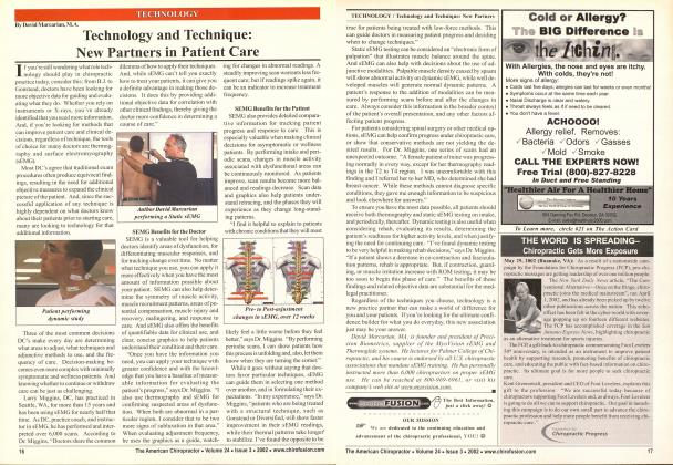
Patients are not only better able to understand the importance of their X-rays but, also, receive the advantage of having the pictures explained to them. h IS INTI-RKSTINCi TO NOT!: THAT, IN Till: last few years, there has been a phenom- enal explosion of cyber information using the ^ internet and the advanced technology worldwide. From Silicon Valley to home transferred garages, computer technology has grown by leaps and bounds. It has also affected, of course, the technology by wliich X-rays are being interpreted for doctors. In the competitive healthcare arena of third party reimbursement, it is crucial to provide the most objective and accurate documentation to support treatment for continued care of each patient. A system called digitization provides such protocol. Indeed, twenty-six different mensurations in all areas of the spine can be clearly defined, using the latest technology. An image is usually acquired either from a direct link to an MRI, CT, or even a scanned existing film, or it can be downloaded via telephone link. Each acquired image is then identified and matched with patient data and stored on the computer system. The system then stores all the basic patients and image information, such as patient name, social security number, film date, image size, image content, referring doctor, etc. Each image is, in turn, analyzed according to its content. Various landmarks arc selected and stored in a location separate from the image. The numbered data points (landmarks) are later used to reconstruct a variety of biomechanical relationships and measurements that are useful to the doctor and the patient. Finally, a comprehensive biomechanical report is generated, using mainstream, widely accepted and conservative mensurations. These twenty-six mensurations are used as per the AMA guide of impairment rating. A wide variety of image types and qualities is obviously delivered. The quality of the image can depend on many factors. While the image is being analyzed, some required landmarks may not be visible, due to the over-exposure or under-exposure of the film. During the analysis process, and while landmarks are being selected, the image enhancements arc available through the latest technologies today. These tools make it possible to view certain parts of the image that were basically unreadable without the use of the computer. After the enhancements are made, they can be reversed and re-applied, so that the best possible view of the total image is obtained. The final enhancements are saved along with the landmarks for later printing. The original digital image is never modified. The separately saved enhancements are simply localized contrast and brightness adjustments (called Histogram Equalization). Whether viewing an original image or an enhanced image, the image biomechanics and structural integrity remain unchanged. Some landmarks may not be made visible by any means, because they are either off-film or obscured. This is often true with the inferior endpoints of C7 on a cervical lateral view, due to the shoulders obscuring the vertebrae. In cases where landmarks are not visible, those measurements that require the landmark arc usually excluded from the reports. Using the latest technology, these pictures are then imprinted on to photographic paper and give a comprehensive biomechani-cal report and digitized analysis. The reports all include impairment ratings per the American Medical Associations Guide to the Evaluation of Permanent Impairment, hence justifying medical necessity. The protocols used by patented software technology are capable of delivering digitized computer analysis, using the actual real pictures, a rare treat to the patient. The patient is able to see his own X-rays which have been digitized and marked with a complete analysis and comprehensive references, showing not only all the mensuration lines but, also, the comparison to normal films. In fact, the patients' images are printed in specialized technological format next to comparatively normal images. The patients are not only better able to understand the importance of their X-rays but, also, receive the advantage of having the pictures explained to them. In conclusion, a pathology report preformed by a radiologist determines the proper findings and the digitized analysis to show computer generated images/pictures of the patients X-rays, demonstrating various measurements with comparative standards, as well as a biomechanical report, showing a test summary of all the findings, including textbook definitions/relationship, and the impairment rating per the AMA guide to the Evaluation of Permanent Impairment (5th Edition). All of this is backed by proper references throughout the reports. Technology is definitely here to stay and the further advances that are constantly being made are necessary to maximize the patients' treatments, and to provide the doctors with the most comprehensive and professional reports available, while limiting their liability and increasing their credibility. EZQ For more information, contact PDl at 866-4PDI-PDI, or visit wxyn.ahvKipdi.com. The best information, just a click a«av! ©