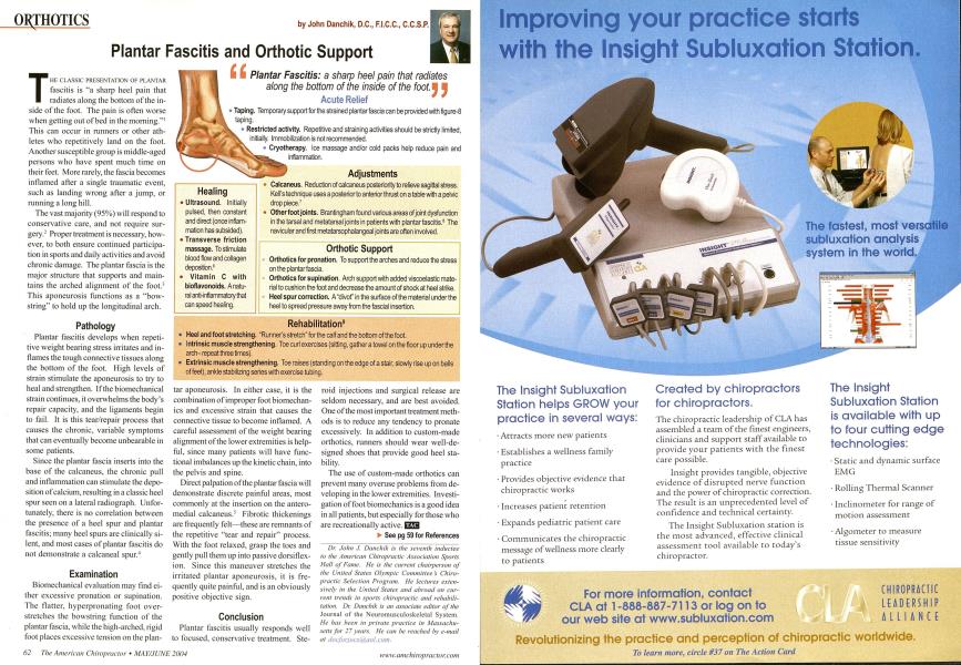
T HE CLASSIC PRESENTATION OF PLANTAR fascitis is "a sharp heel pain that radiates along the bottom of the inside of the foot. The pain is often worse when getting out of bed in the morning."1 This can occur in runners or other athletes who repetitively land on the foot. Another susceptible group is middle-aged persons who have spent much time on their feet. More rarely, the fascia becomes inflamed after a single traumatic event, such as landing wrong after a jump, or running a long hill. The vast majority (95%) will respond to conservative care, and not require surgery.2 Proper treatment is necessary, however, to both ensure continued participation in sports and daily activities and avoid chronic damage. The plantar fascia is the major structure that supports and maintains the arched alignment of the foot.3 This aponeurosis functions as a "bowstring" to hold up the longitudinal arch. Pathology Plantar fascitis develops when repetitive weight bearing stress irritates and inflames the tough connective tissues along the bottom of the foot. High levels of strain stimulate the aponeurosis to try to heal and strengthen. If the biomechanical strain continues, it overwhelms the body's repair capacity, and the ligaments begin to fail. It is this tear/repair process that causes the chronic, variable symptoms that can eventually become unbearable in some patients. Since the plantar fascia inserts into the base of the calcaneus, the chronic pull and inflammation can stimulate the deposition of calcium, resulting in a classic heel spur seen on a lateral radiograph. Unfortunately, there is no correlation between the presence of a heel spur and plantar fascitis: many heel spurs are clinically silent, and most cases of plantar fascitis do not demonstrate a calcaneal spur.4 Examination Biomechanical evaluation may find either excessive pronation or supination. The flatter, hyperpronating foot overstretches the bowstring function of the plantar fascia, while the high-arched, rigid foot places excessive tension on the plan- tar aponeurosis. In either case, it is the combination of improper foot biomechan-ics and excessive strain that causes the connective tissue to become inflamed. A careful assessment of the weight bearing alignment of the lower extremities is helpful, since many patients will have functional imbalances up the kinetic chain, into the pelvis and spine. Direct palpation of the plantar fascia will demonstrate discrete painful areas, most commonly at the insertion on the antero-medial calcaneus/ Fibrotic thickenings are frequently felt—these are remnants of the repetitive "tear and repair" process. With the foot relaxed, grasp the toes and gently pull them up into passive dorsiflex-ion. Since this maneuver stretches the irritated plantar aponeurosis, it is frequently quite painful, and is an obviously positive objective sign. Conclusion Plantar fascitis usually responds well to focused, conservative treatment. Ste- roid injections and surgical release are seldom necessary, and are best avoided. One of the most important treatment methods is to reduce any tendency to pronate excessively. In addition to custom-made orthotics, runners should wear well-designed shoes that provide good heel stability. The use of custom-made orthotics can prevent many overuse problems from developing in the lower extremities. Investigation of foot biomechanics is a good idea in all patients, but especially for those who are recreationally active. EZQ ► See pg 59 for References Dr. John J. Danchik is the seventh inductee to the American Chiropractic Association Sports Hall of Fame. He is the current chairperson of the United Slates Olympic Committee s Chiropractic Selection Program. He lectures extensively in the United States and abroad on current trends in sports chiropractic and rehabilitation. Dr. Danchik is an associate editor of the Journal of the Neuromusculoskeletal System. He has been in private practice in Massachusetts for 27 years. He can be reached by e-mail at docluriocili.iicwl.com. Taping. Temporary support for the strained plantar fascia can be provided with figure-8 taping. Restricted activity. Repetitive and straining activities should be strictly limited, initially. Immobilization is not recommended. Cryotherapy. Ice massage and/or cold packs help reduce pain and inflammation. Healing Ultrasound. Initially pulsed, then constant and direct (once inflam mation has subsided). Transverse friction massage. To stimulate blood flow and collagen deposition.6 • Vitamin C with bioflavonoids. A natu ral anti-inflammatory that can speed healing. Adjustments Calcaneus. Reduction of calcaneus posteriority to relieve sagittal stress. Kell's technique uses a posterior to anterior thrust on a table with a pelvic drop piece.7 Other foot joints. Brantingham found various areas of joint dysfunction in the tarsal and metatarsal joints in patients with plantar fascitis.8 The navicular and first metatarsophalangeal joints are often involved. Orthotic Support Orthotics for pronation. To support the arches and reduce the stress on the plantar fascia. Orthotics for supination. Arch support with added viscoelastic material to cushion the foot and decrease the amount of shock at heel strike. Heel spur correction. A "divot" in the surface of the material under the heel to spread pressure away from the fascial insertion. Rehabilitation9 Heel and foot stretching. "Runner's stretch" for the calf and the bottom of the foot. Intrinsic muscle strengthening. Toe curl exercises (sitting, gather a towel on the floor up under the arch- repeat three times). Extrinsic muscle strengthening. Toe raises (standing on the edge of a stair, slowly rise up on balls of feet), ankle stabilizing series with exercise tubing.