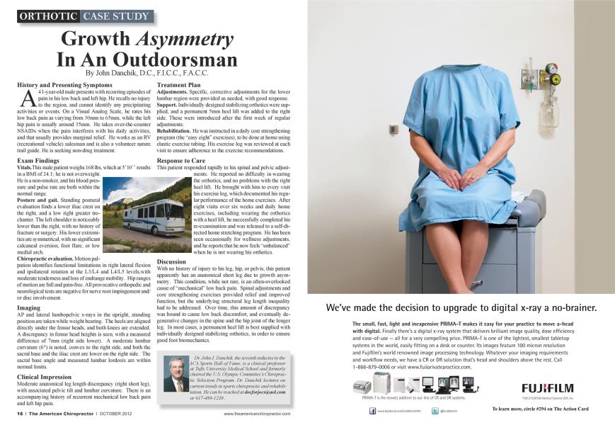
History and Presenting Symptoms A 41 -year-old male presents with recurring episodes of pain in his low back and left hip. He recalls no injury to the region, and cannot identify any precipitating activities or events. On a Visual Analog Scale, he rates his low back pain as varying from 30mm to 65mm. while the left hip pain is usually around 35mm. He takes over-the-counter NSAIDs when the pain interferes with his daily activities, and that usually provides marginal relief. He works as an RV (recreational vehicle) salesman and is also a volunteer nature trail guide. He is seeking non-drug treatment. Exam Findings Vitals.This male patient weighs 168 lbs, which at 5" 10"" results in a BMI of 24.1; he is not overweight. He is a non-smoker, and his blood pressure and pulse rate are both within the normal range. Posture and gait. Standing postural evaluation finds a lower iliac crest on the right, and a low right greater tro-chantcr. The left shoulder is noticeably lower than the right, with no histon of fracture or surgery. His low cr extremities arc symmetrical, with no significant calcancal eversion. foot flare, or low medial arch. Chiropractic evaluation. Motion pal- pation identifies functional limitations in right lateral flexion and ipsilatcral rotation at the L3/L4 and L4/L5 lcvcls.with moderate tenderness and loss of endrange mobility. Hip ranges of motion arc full and pain-free. All provocative orthopedic and neurological tests arc negative for nerve root impingement and/ or disc involvement. Imaging AP and lateral lumbopclvic x-rays in the upright, standing position arc taken while weight bearing. The heels arc aligned directly under the femur heads, and both knees arc extended. A discrepancy in femur head heights is seen, with a measured difference of 7mm (right side lower). A moderate lumbar curvature (6°) is noted, convex to the right side, and both the sacral base and the iliac crest are lower on the right side. The sacral base angle and measured lumbar lordosis are within normal limits. Clinical Impression Moderate anatomical leg length discrepancy (right short leg), with associated pelvic tilt and lumbar curvature. There is an accompanying history of recurrent mechanical low back pain and left hip pain. Treatment Plan Adjustments. Specific, corrective adjustments for the lower lumbar region were provided as needed, with good response. Support. Individually designed stabilizing orthotics were supplied, and a permanent 5mm heel lift was added to the right side. These were introduced after the first week of regular adjustments. Rehabilitation. He was instructed in a daily core strengthening program (the "easy eight" exercises), to be done at home using elastic exercise tubing. His exercise log was reviewed at each visit to ensure adherence to the exercise recommendations. Response to Care This patient responded rapidh to his spinal and pelvic adjust- merits. He reported no diiiiculty in wearing the orthotics. and no problems with the right heel lift. He brought with him to every visit his exercise log. which documented his regular performance of the home exercises. After eight visits over six weeks and daily home exercises, including wearing the orthotics with a heel lift, he suecessfulh completed his re-examination and was released to a self-directed home stretching program. He lias been seen occasionally for wcllncss adjustments, and he reports that he now feels "unbalanced" when he is not wearing his orthotics. Discussion With no history of injury to his leg. hip. or pelvis, this patient apparently has an anatomical short leg due to growth asymmetry. This condition, while not rare, is an often-overlooked cause of "mechanical" low back pain. Spinal adjustments and core strengthening exercises provided relief and improved function, but the underlying structural leg length inequality had to be addressed. Over time, this amount of discrepancy was bound to cause low back discomfort, and eventually degenerative changes in the spine and the hip joint of the longer leg. In most cases, a permanent heel lift is best supplied with individually designed stabilizing orthotics. in order to ensure good foot biomechanics. Dr. John.J. Danchik. the seventh inductee to the ACA Sports Hall of Fame, is a clinical professor at Tufts University Medical School and formerly chaired the U.S. Olympic Committee's Chiropractic Selection Program. Dr. Danchik lectures on current trends in sports chiropractic and rehabilitation. He can he reached at docforjocs(a aol.com or 617-4X9-1220.