Chiropractic, Chronic Back Pain, and Brain Shrinkage
FEATURE
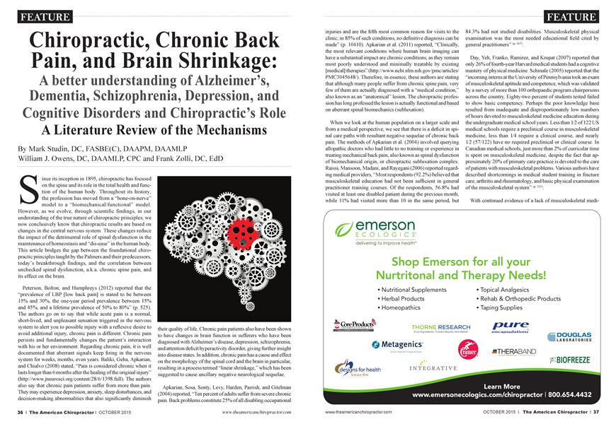
A better understanding of Alzheimer’s, Dementia, Schizophrenia, Depression, and Cognitive Disorders and Chiropractic’s Role A Literature Review of the Mechanisms
Mark Studin
William J. Owens
Frank Zolli
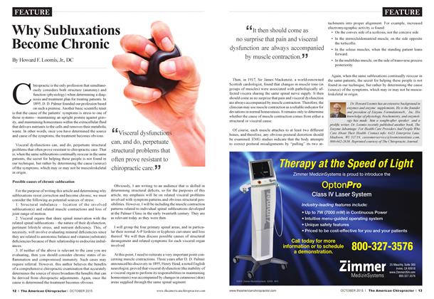
Since its inception in 1895, chiropractic has focused on the spine and its role in the total health and function of the human body. Throughout its history, the profession has moved from a “bone-on-nerve” model to a “biomechanical/functional” model. However, as we evolve, through scientific findings, in our understanding of the true nature of chiropractic principles, we now conclusively know that chiropractic results aie based on changes in the central nervous system. These changes reduce the impact of the detrimental role of spinal dysfunction in the maintenance of homeostasis and “dis-ease” in the human body. This article bridges the gap between the foundational chiropractic principles taught by the Palmers and their predecessors, today’s breakthrough findings, and the correlation between unchecked spinal dysfunction, a.k.a. chronic spine pain, and its effect on the brain.
Peterson, Bolton, and Humphreys (2012) reported that the “prevalence of LBP [low back pain] is stated to be between 15% and 30%, the one-year period prevalence between 15% and 45%, and a lifetime prevalence of 50% to 80%” (p. 525). The authors go on to say that while acute pain is a normal, short-lived, and unpleasant sensation triggered in the nervous system to alert you to possible injury with a reflexive desire to avoid additional injury, chronic pain is different. Chronic pain persists and fundamentally changes the patient’s interaction with his or her environment. Regarding chronic pain, it is well documented that aberrant signals keep firing in the nervous system for weeks, months, even year s. Baliki, Geha, Apkarian, and Chialvo (2008) stated, “Pain is considered chronic when it lasts longer than 6 months after the healing of tire original injury” (http://www.jneurosci.org/content/28/6/1398.full). The authors also say that chr onic pain patients suffer from more than pain. They may experience depression, anxiety, sleep disturbances, and decision-making abnormalities that also significantly diminish
their quality of life. Chronic pain patients also have been shown to have changes in brain function in sufferers who have been diagnosed with Alzheimer’s disease, depression, schizophrenia, and attention deficit hyperactivity disorder, giving further insight into disease states. In addition, chr onic pain has a cause and effect on tire morphology of the spinal cord and the brain in particular, resulting in a process termed “linear' shrinkage,” which has been suggested to cause ancillary negative neurological sequelae.
Apkarian, Sosa, Sonty, Levy, Harden, Parrish, and Gitelman (2004) reported, “Ten percent of adults suffer from severe chronic pain. Back problems constitute 25% of all disabling occupational
injuries and aie the fifth most common reason for visits to the clinic; in 85% of such conditions, no definitive diagnosis can be made” (p. 10410). Apkarian et al. (2011) reported, “Clinically, the most relevant conditions where human brain imaging can have a substantial impact aie cluonic conditions, as they remain most poorly understood and minimally treatable by existing [medical] therapies” (http://www.ncbi.nlm.nih.gov/pmc/articles/ PMC3045648/). Therefore, in essence, these authors aie stating that although many people suffer from chronic spine pain, very few of them aie actually diagnosed with a “medical condition,” also known as an “anatomical” lesion. The chiropractic profession has long professed the lesion is actually functional and based on aberrant spinal biomechanics (subluxation).
When we look at the human population on a larger scale and from a medical perspective, we see that there is a deficit in spinal care paths with resultant negative sequelae of cluonic back pain. The methods of Apkarian et al. (2004) involved querying allopathic doctors who had little to no training or experience in treating mechanical back pain, also known as spinal dysfunction of biomechanical origin, or chiropractic Subluxation complex. Raissi, Mansoon, Madani, and Rayegani (2006) reported regarding medical providers, “Most respondents (92.2%) believed that musculoskeletal education had not been sufficient in general practitioner training courses. Of the respondents, 56.8% had visited at least one disabled patient during the previous month, while 11% had visited more than 10 in the same period, but
84.3% had not studied disabilities. Musculoskeletal physical examination was the most needed educational field cited by general practitioners” (p 167).
Day, Yeh, Franko, Ramirez, and Krapat (2007) reported that only 26% of fourth-year Harvard medical students had a cognitive mastery of physical medicine. Schmale (2005) reported that the “incoming interns at the University of Pennsylvania took an exam of musculoskeletal aptitude and competence, which was validated by a survey of more than 100 orthopaedic program chairpersons across the country. Eighty-two percent of students tested failed to show basic competency. Perhaps the poor knowledge base resulted from inadequate and disproportionately low numbers of hours devoted to musculoskeletal medicine education during the undergraduate medical school years. Less than 1/2 of 122 US medical schools require a preclinical course in musculoskeletal medicine, less than 1/4 require a clinical course, and nearly 1/2 (57/122) have no required preclinical or clinical course. In Canadian medical schools, just more than 2% of curricular time is spent on musculoskeletal medicine, despite the fact that approximately 20% of primary care practice is devoted to the care of patients with musculoskeletal problems. Various authors have described shortcomings in medical student training in fracture care, arthritis and rheumatology, and basic physical examination of the musculoskeletal system” (p-25'/
With continued evidence of a lack of musculoskeletal medi-
cine and a subsequent deficiency of training in spine care, particularly of biomechanical (subluxation) orientation, the question becomes: Which profession has the educational basis, training, and clinical competence to manage these cases? Let’s take a closer look at chiropractic education as a comparison.
According to the American Chiropractic Association, it is fundamental that doctors of chiropractic complete 4,820 hours of training (compared to 3,398 for physical therapy and 4,670 to medicine) and receive a thorough knowledge of anatomy and physiology. As a result, all accredited doctor of chiropractic degree programs focus a significant amount of time in their curricula on these basic science courses. It is so important to practice these courses that the Council on Chiropractic Education, the federally recognized accrediting agency for chiropractic education, requires a curriculum that enables students to be “proficient in neuromusculoskeletal evaluation, treatment and management.” In addition to multiple courses in anatomy and physiology, the typical curriculum in chiropractic education includes physical diagnosis, spinal analysis, biomechanics, orthopedics, and neurology. As a result, students aie afforded the opportunity to practice utilizing this basic science information for many hours prior to beginning clinical services in then internships.
To qualify for licensure, graduates of chiropractic programs must pass a series of examinations administered by the National Board of Chiropractic Examiners (NBCE). Paid one of this series consists of six subjects: general anatomy, spinal anatomy, physiology, chemistry, pathology, and microbiology. Therefore, it is mandatory for a chiropractic school graduate to know the structure and function of the human body to pass this test, and as a result, the study of neuromuscular and biomechanics is woven thr oughout the fabric of chiropractic education. Therefore, the doctor of chiropractic is expert in the same musculoskeletal gerne that medical doctors aie poorly trained in their doctoral education, as evidenced previously.
Now that we have a general idea of why current musculoskeletal and spine care paths aie failing as the majority of the population seeks medical care first, let’s examine what the negative effects aie with a focus on what happens to the central nervous system when a patient is suffering from chronic pain. The following describes what happens to the brain because of chronic pain and then offers solutions based upon evidence-based studies.
Chronic Pain Affecting Brain Activity at Rest
Baliki et al. (2008) reported, “Recent studies have demonstrated that chronic pain harms cortical areas unrelated to pain... long-term pain alters the functional connectivity of cortical regions known to be active at rest, i.e., the components of the ‘default mode network’ (DMN). This DMN is marked by balanced positive and negative correlations between activity in component brain regions. In several disorders, however, this balance is disrupted... Studying with fMRI [functional MRI] a group of chronic back pain (CBP) patients and healthy controls while executing a simple visual attention task, we discovered that chronic back pain patients, despite performing the task equally well as controls, displayed reduced deactivation in several key DMN [default mode network] regions. These findings demonstrate that chronic pain has a widespread impact on overall brain function, and suggest that disruptions of the DMN [default mode network] may underlie the cognitive and behavioral impairments accompanying chronic pain” (http:// www.jneuro sei. org/content/28/6/1398. full).
the existence of a resting state in which the brain remained active in an organized manner... was called the ‘default mode’ of brain function. The regions exhibiting a decrease in activity during task performance are the component members of the ‘defaultmode netM’ork’ (DMN), which in concerted action maintain the brain resting state... Recent studies have already demonstrated that the brain DMN [default mode netM’ork] is disrupted in autism, Alzheimer ’ disease, depression, schizophrenia and attention deficit hyperactivity disorder, suggesting that the study of brain resting activity can be useful to understand disease states as well as potentially provide diagnostic infor-
matiori” (http://www.jneurosci. org/content/2 8/6/1398. full).
This is important since, for the first time, we are starting to see a published correlation between spinal function, chronic pain, and central nervous system changes. This is what our chiropractic founders observed yet were unable to prove.
“Thus, the alterations in the patient’s brain at ‘rest’ can result in a different DMN [default mode network] organization. In turn, potential changes in the default-mode network activity could be related to symptoms (other than pain) commonly exhibited by chronic pain patients, including depression and anxiety, sleep disturbances, and decision-making abnormalities, which also significantly diminish their quality of life... chronic pain patients display a dramatic alteration in several key default-mode network regions, suggesting that chronic pain has a widespread impact on overall brain function” (http://www.jneurosci. org/content/28/6/1398. full). This information points to the fact that a doctor of chiropractic should be involved in the triage and treatment of these patients and part of a long-term spinal care program based upon the evidence in the conclusion.
Baliki et al. (2008) continued, “Consistent with extensive earlier work examining visuospatial attention tasks, dominant activations were located in posterior parietal and lateral prefrontal
^It seems that enduring pain for a long time affects brain function in response to even minimally demanding attention tasks completely unrelated to pain. ï Ï
cortices, whereas deactivations occurred mainly within mPFC [prefrontal cortex] and PCC [posterior cingulate/cuneate cortexes]. Although activations in CBP [chronic back pain] patients’ and controls’ brains were similar, CBP exhibited significantly less deactivations than healthy subjects in mPFC [prefrontal cortex], amygdala, and PCC [posterior cingulate/ cuneate cortexes]... The focus was on identifying differences in the way CBP patients’ brains process information not related to pain. This is the first study demonstrating that chronic back pain patients exhibit severe alterations in the functional connectivity between brain regions implicated in the default mode network. It seems that enduring pain for a long time affects brain function in response to even minimally demanding attention tasks completely unrelated to pain. Furthermore, the fact that the observed task performance, compared with healthy subjects, is unaffected, whereas the brain activity is dramatically different, raises the question of how other behaviors aie impaired by the altered brain activity’’ (http://www.jneurosei.org/content/28/6/1398.full).
However, the disruption of functional connectivity observed here with increased chr onic back pain duration may be related to file earlier observation of bram atrophy increasing with pain duration also in chronic back patients. CBP patients exhibit increased mPFC [prefrontal cortex] activity in relation to spontaneous
pain, in addition to dorsolateral prefrontal cortex (dIPFC) atrophy. Therefore, the decreased deactivations described here may be related to the dIPFC [dorsolateral prefrontal cortex]/mPFC [prefrontal cortex] mutual inhibitory interactions perturbed with time. If that is the case, it will support the idea of a plastic, timedependent, reorganization of the brain as patients continue to suffer from chr onic back pain.
“Mechanistically, the early stages of this cortical reorganization may be driven by peripheral and spinal cord events, such as those that have been documented in animal models of chronic pain, whereas later events may be related to coping strategies necessary for living with unrelenting pain. It is important to recognize that transient but repetitive functional alterations can lead to more permanent changes. Accordingly, long-term interference with normal default mode network activity may eventually initiate plastic changes that could alter irreversibly the stability and subsequently the conformation of the resting state networks” (http : //www.j neuro sci. org/content/28/6/1398.full).
Chronic Pain Causing Brain “Shrinkage”
Apkarian et al. (2004) reported, “CBP [chronic back pain] patients were divided into neuropathic, exhibiting pain because of sciatic nerve damage, and non-neuropathic groups... Patients with chronic back pain showed 5-11% less neocortical gray matter volume than control subjects. The magnitude of this decrease is equivalent to the gray matter volume lost in 10-20 years of normal aging. The decreased volume was related to pain duration, indicating a 1.3 cm3 loss of gray matter for ev-
ery year of chronic pain... Gray matter density was reduced in bilateral dorsolateral prefrontal cortex and right thalamus and was strongly related to pain characteristics in a pattem distinct for neuropathic and non-neuropathic chronic back pain. Our results imply that chronic back pain is accompanied by brain atrophy and suggest that the pathophysiology of chr onic pain includes thalamocortical processes” (p 10410)
“it is assumed that the cerebral cortex passively reflects spinal changes and reverts to its normal state after cessation of chr onic pain. Our studies show that chronic back pain (CBP) (sustained for >6 months) is accompanied by abnormal brain chemistry, mainly a reduction in the N-acetylaspartate-creatine ratio in the prefrontal cortex, implying neuronal loss or dysfunction in this region and reduced cognitive abilities on a task that implies abnormal prefrontal processing” (APkanan et al> 2004> P1041°)
Apkarian et al. (2004) continued, “At the whole-brain level, this reduction is related to pain duration, regionally depends on multiple pain-related characteristics, and is more severe in the neuropathic subtype. Therefore, these data present strong evidence that the pathophysiology of chronic pain includes cortical processes, and the observed changes likely constitute the physical substrate of the cognitive and behavioral properties of chr onic pain” (p 104114
“Thus, regional gray matter changes aie strongly and specifically related to pain characteristics, and this pattern is opposite for neuropathic compared with non-neuropathic types. This dissociation is consistent with extensive clinical data showing
that neuropathie pain conditions are more debilitating and have a stronger negative effect, which may be directly attributable to the larger decrease in gray matter density that we observe in the DLPFC [dorsolateral prefrontal cortex] of nuCBP [neuropathic chronic back pain] patients”(Apkanan et aL 2004-p 10413)
“In the DLPFC, a larger proportion of the variance could be explained by pain characteristics (40% for nuCBP [neuropathic chronic back pain]; 80% for nonneuropathic chronic back pain), implying a tighter relationship between regional brain atrophy and perceived pain. Therefore, we suggest that the pattem of brain atrophy is directly related to the perceptual and behavioral properties of neuropathic chronic back pain”
(Apkarian et al., 2004, p. 10413)
“The observed regional pattem of atrophy is distinct from that seen in chronic depression or anxiety and shows a minimal relationship with anxiety and depression traits. Thus, it seems to be specific to chronic pain, especially because the regions showing atrophy, the thalamus and DLPFC, participate in pain perception. The DLPFC is activated in acute pain, with responses that do not code stimulus intensity. Recent evidence suggests that the DLPFC exerts ‘top-down’ inhibition on orbi-
^Thalamic atrophy in chronic back pain is important because it is a major source of nociceptive inputs to the cortex J J
tofiontal activity, limiting the magnitude of perceived pain. Thus, DLPFC [dorsolateral prefrontal cortex] atrophy may lead to a disruption of its control over orbitofiontal activity, which in turn is critical in the perception of negative effect in general and particularly in pain states. Thalamic atrophy in chronic back pain is important because it is a major source of nociceptive inputs to the cortex and damage to this region may be a reason for the generalized sensory abnormalities commonly associated with chronic pain”
(Apkarian et al., 2004, p. 10413).
“The dorsal anterior cingulate is shown to be specifically involved in pain affect in normal subjects and exhibits decreased nociceptive signaling in various chronic pain states, which may again be caused by thalamic atrophy because the anterior thalamus is a primary input to the anterior cingulate. Therefore, we suggest that regional atrophy dictates the brain activity observed in chronic pain, and it may explain the transition from acute to chronic pain by shifting brain activity related to pain affect away from the anterior cingulate to orbitofiontal
(Apkarian et al., 2004, p. 10414).
“It is possible that some of the observed decreased gray matter reflects tissue shrinkage (changes in extracellular space and microvascular volume may cause tissue shrinkage without substantially impacting neuronal properties), implying that proper treatment would (authors’ note: could instead of would) reverse this portion of the decreased brain gray matter. The atrophy may be also attributable to more irreversible processes, such as neurodegeneration, which we favor because the main brain region involved (the DLPFC) also exhibits decreased N-acetylaspartate, and decreased N-acetylaspartate has been observed in most neurodegenerative conditions... Recent evidence also suggests that after nerve injury, some components of pain behavior aie a consequence of hyperactivity of spinal cord microglia, and a histological study has shown a reduction in glial numbers in the cortex in major depressive disorder and bipolar
disorder” (Apkarian et al., 2004, p. 10414).
This article suggests that there is a reversible component in brain atrophy with the resolution of the chronic back pain since there is strong evidence that there are some tissue structures that will be permanently damaged should the chr onic pain go beyond the defined six months.
Clearly, there are many different professions that handle the anatomical components of spine pain, such as fracture, infection, disc herniation, or tumor. There is only one profession that has the education and training to treat the aberrant spinal biomechanics—chiropractic. Since chiropractors are trained in treating, managing, and triaging the anatomical lesions while also being the best suited to treat the biomechanical component, the evidence verifies that the first contact for spine pain should be a doctor of chiropractic who is also trained in differential diagnosis of underlying pathology.
Brain Regions Affected
Apkarian, Hashmi, and Baliki (2011) reported, “The surprise was that the brain region best reflecting high magnitude of back pain was localized to the medial prefrontal cortex (mPFC), extending into anterior ACC [cingulate cortex]; a region not anticipated by acute pain studies. Additionally, brain areas observed for acute pain, like portions of the insula and mid-ACC [anterior cingulate cortex] were only active transiently and only when the back pain magnitude was on the increase. These results aie exciting because for the first time we are able to observe brain activity reflecting the subjective perception of the pain that chr onic back pain patients come to the clinic to complain about.. .We interpret the transient activity as a nociceptive signal from the periphery, which then is converted into a sustained emotional suffering signal in mPFC [medial prefrontal cortex]” (http : //www. ncbi. nlm. nih. go v/pmc/article s/PMC3045 648/).
“Thus we can assert that, at least in this group of chronic pain patients, different brain areas encode the perceived magnitude for distinct types of pain. The prevalent expectation for brain activity in chronic pain is a sustained or enhanced activation of the brain areas already identified for acute pain. This view is partly implied by the chronic pain definition and by notions of specificity theory or labeled line theory of pain (where supraspinal organization and representation of pain is assumed to be through fixed and immutable routes). This is exactly what we do not see. Instead these results imply that functional anatomy or physiology or some combination of both have changed in the brain of chronic back pain patients” (APkananetal -2011http //www
ncbi.nlm.nih.gov/pmc/articles/PMC3045648/).
“It is also important to remember that the close relationships between fundamental properties of back pain and activity in mPFC [medial prefrontal cortex] and insula are correlational, and both mPFC and insula respond to a long list of cognitive and emotional states...The morphological studies show that the brain structure undergoes changes at multiple spatial and temporal scales, which are for the most part specific to the type of chronic pain studied. That some of these changes are
reversible by cessation of chronic pain speaks to the specificity of the processes and also demonstrate that chronic pain may in fact by used as a unique tool with which the dynamics of brain plasticity can be studied at multiple spatial and temporal scales”
(Apkarian et al., 2011, http://www.ncbi.nlm.nih.gov/pmc/articles/PMC3045648/).
Chiropractic as a Solution for Chronic Back Pain
Peterson et al. (2012) set out to “investigate outcomes and prognostic factors in patients with acute or chronic low back pain (LBP) undergoing chiropractic treatment” (p. 525). They made note, “In chr onic LBP, recent studies indicate that significant improvement is often fairly rapid, usually by the fourth visit, and that patients initially receiving treatment thr ee to four
times a week have better outcomes” (Peterson et ah, 2012, p. 526). They discovered, “Patients with chronic and acute back pain both reported good outcomes, and most patients with radiculopathy [neurogenic] also improved”
(Peterson et al., 2012, p. 525). tllTCC
■ "This is exactly what we do not see. Instead these results imply that functional anatomy or physiology or some combination of both have changed in the brain of chronic back pain patients J J
months... 69% of patients with chronic pain stated that they were either much better or better” (Peterson et ah, 2012, p. 528). “This is unlikely to be due to the natural history of LBP because these patients have already passed the period when natural history occurs” (Peterson et ah, 2012, p. 531). A study by Tamcan, Mannion, Eisenring, Horisberger, Elfering, and Müller (2010) was the only population-based study regarding the so-called “natural history” of low back pain, and the authors found the “natural history” of chronic lower back pain was not ending in a resolution of symptoms. Instead, they documented patients moving “in and out” of a level of pain they could tolerate. Based on this, the only population-based study of chronic lower back pain, the idea that the “natural history” of low back pain ends with resolution of symptoms is a complete myth and one that is perpetuated by our present healthcare system.
Lawrence et al. (2008) reported, “Existing research evidence regarding the usefulness of spinal adjusting...indicates the following:
1. As much or more evidence exists for the use of SMT [spinal manipulative therapy, i.e., chiropractic spinal adjustment] to reduce symptoms and improve function in patients with chr onic low back pain as for use in acute and subacute LBP” (p. 670).
“...the manual therapy group showed significantly greater improvements than did the exercise group for all outcomes. Results were consistent for both the short term and the long
tCim” (Lawrence et al., 2008, p. 663).
Dunn, Green, Formolo, and Chicoine (2011) reported, “The clinical outcomes achieved for this sample should be considered within the context of this veteran patient base, which is typically represented by older, white males with multiple comorbidities. A high percentage of overall service-connected disability was
noted, with only a small percentage associated with the low back region. Considerable psychological comorbidity was found, with a high prevalence of PTSD [post-traumatic stress disorder] and depression diagnoses. PTSD and chronic pain tend to co-occur and may interact in a way that can negatively affect either disorder. A previous retrospective study of chiropractic management for neck and back pain demonstrated less improvement among those with PTSD. These points are significant because severe comorbidities and psychosocial factors lessen the likelihood of obtaining positive outcomes with conservative measures, including [chiropractic adjustments], for chronic LBP [low back pain]...,” (p. 930). The authors went on to conclude that in spite of significant comorbidities that have historically compromised positive results, 60.2% of patients met or exceeded the minimum clinically important difference for improvement.
Conclusion
Chronic pain is defined by that which has lasted for six months or longer that causes significant brain aberration in both morphology (size) and function. The literature suggests that this could be the precursor for many diseases as sequelae of the human body’s natural reaction to prolonged pain. Chronic back pain is one of the leading causes of chronic pain and medicine has little to no training on how to manage it or resolve it as reported in the literature. Conversely, chiropractic has significant training and has been proven in “blind” studies to have
significant positive outcomes, even in significantly adverse conditions, to help resolve chronic pain. As a result, the negative sequelae on the brain of a patient with chronic pain, including shrinkage of the brain, can be reversed thr ough chiropractic care by treating the cause (the neuromuscular-mechanical lesion). The evidence has verified that once the chronic pain has resolved, the brain has the ability to return to its normal size and regain much function.
Although this evidence is strong, more research is needed, and this further sets the foundation for understanding how chiropractic directly affects diseases in the human body. In addition, this also takes the chiropractic profession to the next level of understanding regarding how and why a chiropractic adjustment works.
References
1. Peterson, C. K., Bolton, J., & Humphreys, B. K. (2012). Predictors of improvement in patients with acute and chronic low back pain undergoing chiropractic treatment. Journal of Manipulative and Physiological Therapeutics, 35(7), 525-533.
2. Baliki, M. N., Geha, P. Y, Apkarian, A. Y, & Chialvo, D. R. (2008). Beyond feeling: Chronic pain hurts the brain, disrupting the default-mode network dynamics. Journal of Neurosciences, 28(6) http : //www.j neurosci. org/content/28/6/13 98. full
3. Apkarian, Y, Sosa, Y, Sonty, S., Levy, R., Harden, N., Parrish, T, & Gitelman, D. (2004). Chronic back pain is associated with decreased prefrontal and thalamic gray matter density. The Journal of Neuroscience, 24(46), 10410-10415.
4. Apkarian, A. V, Hashmi, J. A., & Baliki, M.N. (2011). Pain and the brain: Specificity and plasticity of the brain in clinical chronic pain. Pain, 152(Suppl. 3). Retrieved from http://www.ncbi.nlm. nih.gov/ pmc/articles/PMC3045648/
5. Raissi, G. R., Mansoon, K., Madani, P, & Rayegani, S. M. (2006). Survey of general practitioners’ attitudes toward physical medicine and rehabilitation. International Journal of Réhabilitât on Research, 29(2), 167-170.
6. Day, C. S., Yeh, A. C., Franko, O., Ramirez, M., & Krupat, E. (2007). Musculoskeletal medicine: An assessment of the attitudes of medical students at Harvard Medical School. Academic Medicine, 82(5), 452-457.
7. Schmale, G. A. (2005). More evidence of educational inadequacies in musculoskeletal medicine. Clinical Orthopaedics and Related Research, 437, 251-259.
8. Tamcan, O., Mannion, A. F., Eisenring, C., Horisberger, B., Elfering, A., & Müller, U. (2010). The course of chronic and recurrent low back pain in the general population. Pain, 150(3), 451-457.
9. Lawrence, D. J., Meeker, W., Branson, R., Bronfort, G., Cates, J. R., Haas, M.,.. .Hawk, C. (2008). Chiropractic management of low back pain and low back-related leg complaints: A literature synthesis. Journal of Manipulative and Physiological Therapeutics, 31(9), 659-674.
10. Dunn, A. S., Green, B. N., Formolo, L. R., & Chicoine, D. (2011). Retrospective case series of clinical outcomes associated with chiropractic management for veterans with low back pain. Journal of Rehabilitation Research & Development, 48(8), 927-934.
Dr. Mark Studin is an adjunct associate professor of ' jL chiropractic at the University of Bridgeport College Of ~ j JÊ Chiropractic and a clinical presenter for the State of He Æ New York at Buffalo, School of Medicine and Biomedical W . V Sciences for postdoctoral education, teaching MRI spine \ AH interpretation and triaging trauma cases. He is an Adjunct Professor, Division of Clinical Sciences, Texas Chiropractic College and also the president ofthe Academy ofChiropractic, teaching doctors of chiropractic how to interface with the legal community (www. DoctorsPIProgram.com). He teaches MRI interpretation and triaging trauma cases to doctors of all disciplines nationally, and studies trends in health care on a national scale (www. TeachDoctors.com). He can be reached at DrMarkiaA cade my of( 'hiropractic. com or at 631-786-4253.
Dr Bill Owens is presently in private practice in Buffalo and Rochester, New York, and generates the majority of his new patient referrals directly from the primary care medical community. He is an associate adjunct professor at the State HI University of New York at Buffalo, School of Medicine and Biomedical Sciences as well as the University of Bridgeport, College of Chiropractic. He is an Adjunct Professor, Division of Clinical Sciences, Texas Chiropractic College and works directly with doctors of chiropractic to help them build relationships with medical providers in their community. He can be reached by e-mail at drowens a academyofchiropractic.com, through www.mdreferralprogram.com, or by calling716-228-3847.
Dr Frank Zolli is the founding Dean of the University W J of Bridgeport College of Chiropractic. He is currently a T M professor of clinical sciences at UBCC teaching Orthopedic and Neurologic Testing, Ethics, Jurisprudence and Risk management and the History and Philosophy ofChirpractic. Dr Zolli also maintains a private practice in Orange, Connecticut He can be reached at zolliIjbridgeport.edu or 203 576-4434.