Coccyx Pain and Treatment
TECHNIQUE
Part Two
Continued from Part 1, Issue #3
Jeffrey Tucker
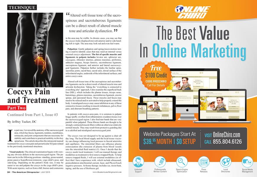
In part one, I reviewed the anatomy of the sacrococcygeal area, which has fascia, ligaments, tendons, membranes, retinacula, and joint components that are necessary for stability and contribute to practical mobility in this key spinal area. This article describes the intrarectal manual treatment for coccyx area pain and periarticular SI J pain related to the previously mentioned structures.
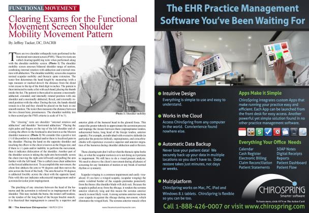
Visual analysis: The clinical examination begins with viewing any obvious defects in the sacrococcygeal region. The patient can be in the following positions: standing, prone neutral, prone passive hyperflexion/extension, yoga child’s pose, and side-lying. Depending on the patient’s body size, it may be easier to see and palpate the coccyx in the yoga child’s pose. With acute injuries, such as from a fall, bruises and contusions
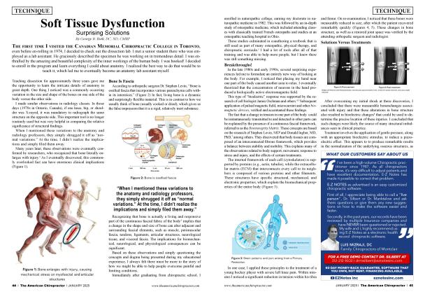
■ ^Altered soft tissue tone of the sacrospinous and sacrotuberous ligaments can be a direct result of altered muscle tone and articular dysfunction. * ?
in the area may be visible. In chronic cases, you may see that the coccyx looks displaced (moved) anterior and/or side bending left or right. The area may look red and even feel wann.
Palpation: Gentle palpation and spring/micro-motion testing is used to identify cases that may need an internal versus external coccyx adjustment. The list of specific muscles and ligaments to palpate includes levator ani, sphincter ani, coccygeus, obturator intemus, gluteus maximus, piriformis, adductor magnus, biceps femoris, sacrotuberous ligament, sacrospinous ligament, and anterior and lateral sacrococcygeal ligaments. Palpation further includes the lumbar spine, sacroiliac joints, sacral base, sacral sulci, dorsal surface of the inferolateral angles, underside of the inferolateral surfaces, and entire coccyx area.
Altered soft tissue tone of the sacrospinous and sacrotuberous ligaments can be a direct result of altered muscle tone and articular dysfunction. Taking the "everything is connected to eveiything else” approach, I also examine the superficial back line (SBL), which includes the plantar tissues, triceps surae, hamstrings, gluteus maximus, sacrotuberous ligament, erector spinae, and epicranial fascia. These muscles and fascia may need to be relaxed and/or activated to help properly extend the body. A misaligned coccyx may cause inhibition in any of these connective tissues resulting in muscle imbalances, pelvic floor pain, and aberrant breathing patterns.
In patients with coccyx-area pain, it is common to palpate boggy (puffy; swollen from inflammatory exudates) tissue over the sacrococcygeal region. I also find taut bands that are very painful when palpated. These fibrous bands are thought to be strongly contracted muscle fibers within an otherwise relatively normal muscle. They may result from protective guarding due to a calcified and misaligned sacrococcygeal joint.
The coccyx was not designed to be up against a chair all day long. The local blood supply and the local tissue viscosity can change influencing local pressure in fascial arterioles and capillaries. The interstitial fibers can influence plasma extravasation (the extrusion of plasma from blood vessels into the interstitial fluid matrix) (1). Once I decide that the coccyx needs local treatment, I will use manual therapy and modalities as treatment to first remove trapped fluids. To help remove trapped fluids, I will use external modalities (in office) that I have experience with, which include ultrasound, premodulation interferential current, laser, and PhysioTouch negative pressure. At-home therapy includes ice, moist heat, taping, and the use of Biofreeze gel.
Treatment: I begin the process of removing the external trapped fluids, (PhysioTouch technique) pulling the attached soft tissues in a posterior direction (I pick several areas where I can pull the tissue posterior). After holding for a period of 30 to 60 seconds, the tissues surrounding the coccyx should begin to release. A contract-relax method can be used in conjunction with this technique. As the coccyx is pulled posterior, the patient is asked to do a gentle contraction of the pelvic floor muscles for five to eight seconds. Upon relaxation, the coccyx can be moved further posterior.
Depending on my results from the external treatment(s), history, mechanism of injury, position of the coccyx, and palpation (tenderness, joint adhesions, mobility restrictions, fusing, etc.), I may need to perform an intrarectal procedure. This is done to drain internally trapped fluids, relax internal soft tissues, restore joint motion, and promote (reintroduce) the healing process.
Intrarectal treatment: Before performing any type of coccyx work, take time to clearly explain what you’re doing and the desired outcome. I inform patients of the procedure, get their consent, and ask them if they would like another person in the room. For obvious reasons, you need gloves and K-Y Jelly. I typically have the patient in a prone or side-lying position.
The specific localized manual intrarectal treatment: Decide on patient position—lying face down, side-lying, or on all fours. The doctor inserts a gloved finger with K-Y Jelly into the rectum of the patient. The internal finger is inserted following the axis of the rectum. Assess the pelvic muscle tone. Slowly and gradually push upward (posteriorly), stretching the pelvic floor, until contact is made with the coccyx, whereupon the pull is released. If the finger is returned immediately to its initial position in the rectum by the patient’s muscle tone without any conscious effort by the patient, the muscle tone is considered abnormally high. Gently and slowly motion-palpate the coccyx in multiplanar directions rather than to actually “move” or manipulate the coccyx. I see the best results with stretching the muscles attached to the sacrococcyx and massaging the ligaments, without actually “moving” the coccyx. Massage in the direction of the fibers.
Gentle coccygeal mobilization: Keep the coccyx in hyperextension while the patient takes several deep breaths; this stresses the sacrococcygeal and intercoccygeal joints and stretches the levator anus. Massage under the lateral edges of the side-bent coccyx, attempting to release the tissue binding the misaligned joint.
Patient instructions: The typical RICE acronym (rest, ice, compress, and elevate) is not the most effective tool for this type of case. An acronym published in the British Journal of Sports Medicine (2) is the POLICE guideline, which exchanges “rest” for “optimal loading.” It is important to protect this particular painful site with a pillow or donut cushion while sitting, but we can’t have the patient sitting “more” as part of the rest! For “optimal loading,” I encourage the patient to get up every
"The coccyx was not designed to be up against a chair all day long. J J
20 minutes, walk two minutes or 20 yards, and look out the window at least 20 yards (20-20-20 tool). Because the real estate of the coccyx is such a small area, I recommend about 10 minutes of ice (cold pack) over the area. Bioffeeze gel is helpful as well. For the “E” in POLICE, rather than elevation, I think of ergonomics—a standing desk (HumanScale.com) and ergonomic chair may be suggested. For a sleeping position (in a right-hand dominant person), I usually recommend if you sleep on your left side, place a pillow under your waist and one or two pillows between the knees. If you sleep on the right side, place a pillow under the waist and one or two pillows between the ankles. One recurring theme seems to be that movement is essential—manually achieving motion.
The treatment process includes checking and balancing the glut max, bicep femoris, lumbodorsal fascia, and piriformis muscles. The patient may need specific stretches and strengthening exercises. These muscles are in good position to maintain balance of the sacral area. It is also helpful to teach coccydynia sufferers a “reverse Kegel” maneuver that fires the pelvic floor muscles, which pushes on the ligamentous web encasing the coccyx.
Discussion: I start with external manual therapy to balance musculofascial tissues around the sacrococcygeal region, pelvic bowl, and lumbar spine. Some patients may need intrarectal coccyx therapy to restore coccyx alignment, reduce inflammation, and promote circulation for drainage of fluids and metabolites.
After the examination and best practices decision-making process, I talk to the patient about treatment options. It’s not likely that I will perform the internal coccyx procedure on the first visit. I like to see how the external treatment goes. My usual procedure is to let the patient know that, on the next office visit, I may perform the intrarectal maneuver. I explain the procedure ahead of time to give the patient a chance to respond from the external procedures. I always ask the patient’s permission to perform the intrarectal technique because it is a hypersensitive and painful area.
References:
1. Kruger, L. (1987). Cutaneous sensory System. In G. Adelman (Ed.), Encyclopedia of Neuroscience (p. 293). Boston, MA: Birkhauser
2. BJ Sports Med, 2012, 6 (4), 220-221
3. Comparison of Three Manual Coccydynia Treatments: A Pilot Study Spine, 2001, 26 (20), E479-E484 JeanYvesMaigne, MD, and Gilles Chatellier, MD
of the Year Jeffrey 2012, Tucker and is the the current ACA Rehabilitation ACA Rehab Council Council secretary/ Doctor treasurer He practices in west Los Angeles, California. His website is www. DrJeffrey Tucker com