Motion X-ray in the Chiropractic Practice
FEATURE
Dennis Woggon
When I first started practicing in 1974, I would get up early, sit down in a quiet place, close my eyes, and visualize the normal spine. I would then, in my mind, subluxate and misalign the spine, and then correct it. This was based on the book Psycho-Cybernetics by Maxwell Maltz. It allowed me to appreciate the art of visualization.
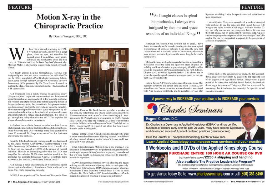
As I taught classes in spinal biomechanics, I always was intrigued by the time and space restraints of an individual Xray. In 1992,1 completed a Post Graduate Continuing Education Course through Palmer College of Chiropractic in video fluoroscopy (VF), presented by Dr. Vem Pierce. I started to explore the idea of the spine in motion, just as I had visualized it 18 years earlier.
As I progressed from a family practice to a personal injury (PI) practice, then I began to focus on scoliosis and the abnormal spinal biomechanics associated with it. For example, we know that rotation and lateral flexion are a normal coupling motion in the upper thoracic spine, but in scoliosis, the spinouses rotate into the concavity and not the convexity, as would be expected. These abnormal spinal biomechanics could be explained by adverse mechanical traction on the spinal cord, which would cause abnormal rotation to reduce the adverse tension. It’s easier to go “through the valley than over the hill.”11 This explains the abnormal coupling motion in scolisois.
As I delved into scoliosis, I started to realize how little doctors of chiropractic and medical doctors understood about scoliosis. I was blessed to have Dr. Fred Barge as my field doctor when I was 16 years old. Dr. Barge wrote one of the few books on chiropractic and scoliosis.
I met Dr. John Postlethwaite years ago and I was fascinated by his Digital Motion X-ray (DMX) system because it was video fluoroscopy (VF) taken to another level. It would take 30 X-rays in one second at 1/1000 of the amount of normal radiation. There is an intensifying tube with the DMX that magnifies the radiation 28,000 times and then sends it to the computer. For example, for regular X-rays, I would take them at 100 mA, but the DMX would take them at 2 mA.m
As I pursued a better understanding of the abnormal spinal biomechanics of scoliosis, I found some DMX studies of scoliosis. This really piqued my curiosity.
In 2006,1 was a speaker at The American Chiropractic Con-
vention in Panama. Dr. Postlethwaite was also a speaker. At that time, my wife Brenda and I had a family cabin in northern Wisconsin that we took care of so others could enjoy it. After listening to Dr. Postlethwaite’s presentation on DMX, Brenda said, “Dennis, you need one of these DMX units to understand scoliosis. Sell the cabin and buy one of these.” So I did, and in 2007,1 bought my DMX system. I will admit that I use it more than the cabin in Wisconsin.
Before I got the Motion X-ray, I considered myself to be great at spinal manual and instrument adjusting because I would base all of my manual adjusting on the patient’s X-rays, and I saw good post-X-ray changes.
When I started utilizing Motion X-ray in my practice, I was amazed at the fact that 99% of my patients had ligament laxity resulting in hypennobility of vertebral segments. I remembered that we were taught in chiropractic college not to adjust hypennobile segments.
In 2007,1 discontinued manual cervical adjusting and began utilizing specific instrument adjusting of the cervical spine only. At this time, I started doing DMX studies on various adjusting instruments. I have found the ArthroStim at 6 Hz to be most effective. Dr. Chris Colloca, DC, found that 6 Hz or 167 milliseconds were most effective in osseous adjustments.1'
“Asi taught classes in spinal biomechanics, I always was intrigued by the time and space restraints of an individual X-ray. J Ï
Although the Motion X-ray is useful for PI cases, I have found it extremely useful in understanding the abnormal spinal biomechanics of scoliosis patients. I can honestly state that I can understand a scoliotic spine in 30 seconds, whereas it took me two weeks to figure out the same thing before with static x-rays.
Motion X-ray as well as flexion and extension x-rays allows the Doctor to see the spine and figure out areas of spinal instability and loss of motion segment integrity (LMSI - AMA Guide to Evaluation of Permanent Impairment, Fifth Edition, Chapter 15v), as well as ligament laxity". This allows me to prescribe specific spinal isometric exercises based on the patient’s X-rays and needs.
Lateral flexion A-P Open Mouth x-rays allows one to see Alai' ligament instability at a certain time and place. Motion X-ray also allows the Doctor to see the abnormal motion associated with Alar ligament instability and to correlate cervical alar
ligament instability™ with the specific cervical spinal instrument adjustment.
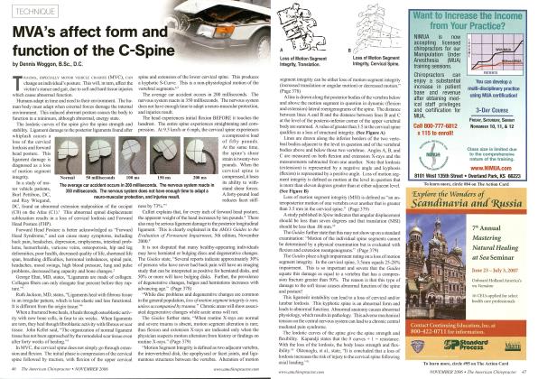
Lateral flexion X-rays are considered a medical standard with scoliosis to see the reduction that lateral flexion will bring to the spine into the Cobb angle. With the Motion Xray, not only can we see the changes in lateral flexion into the Cobb angle, but, by going into the opposite side, we also can see the prognosis and potential for worsening of the Cobb angles. This is very important in regards to the prognosis of scoliosis progression.
In this study of the cervical-dorsal angle, the left cervicaldorsal angle decreases from 12 degrees to the opposite side of -14 degrees with right lateral flexion. This presents a good potential for correction. With left lateral flexion, the cervicaldorsal angle increases to 27 degrees. This has a potential for worsening, but it indicates the necessity for specific spinal isometric exercises.
In the motion study with lateral flexion to the right, the thoracic Cobb angle decreases from 18 degrees to 0 degrees. While this is a good reduction, it is not normal spinal biomechanics since it should flex to the opposite side. The left lateral flexion should increase the thoracic Cobb angle, but in these circumstances, it stays the same.
In the low back, the lumbodorsal Cobb angle decreases with left lateral flexion from 27 degrees to 1 degree, which demonstrates good flexibility. This also reveals a poor prognosis unless stability is addressed.
The DMX allows me to take instant pictures during the study to see where the spine is in time and space. With X-rays and MRIs, you actually can’t be certain that the patient is moving or even alive if you simply review the images or the report. The DMX has picture-in-picture technology to see the patient in time and space.
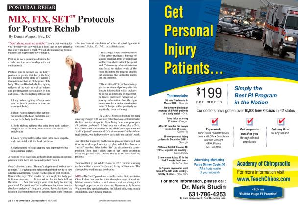
The CLEAR scoliosis protocols also require the scoliosis traction chair. Scoliosis patients aie positioned in the scoliosis traction chair and then a DMX study is done to verify correct placement followed by an X-ray. This is a valuable tool because
an incorrect position will not benefit the patient. A standard 14 x 17 scoliosis X-ray emits 15 times less radiation than a full-spine X-ray. A DMX scoliosis study reduces the radiation exposure by 92%.1
References
z'. http://www.amazon.com/Psycho-Cybernetics-New-MoreLiving-Life dp/0671700758/ref=sr 11 ?ie=UTF8&qid=1425 772275&sr =8-l&keyw ords =psycocyberne tics 77. http://www. clear-institute. org/docs/ScoliosisCC.pdf Hi. http://www. ideal spine, com pages/ajeeJuly 06 instrument adjustingsmechanical advantage.htm
^ * With X-rays and MRls, you actually can’t be certain that the patient is moving or even alive if you simply review the images or the report. 5 5
7v. http://www.ncbi.nlm.nih.gov/pmc/articles/PMC1482708/
v. http://www. amazon. com/Guides-Evaluation-PermanentImpairmenwww. dmxworks. comt-Fifth/dp/15 794 70858/ref=sr
1 _2?s=books&ie=UTF8&qid=1425773954&sr=l-2&keyw ords=ama+guides+to+impairment
vi. http://www. spinalkinetics. info/QA march 2012.pdf
vii. http://www. theamericanchiropractor. com Alar Ligament Laxity.
Dr. Dermis Woggon is the founder of the CLEAR Scoliosis Institute and the St. Cloud Chiropractic Clinic. He graduated from Palmer College of Chiropractic in 1974. He is an international instructor for CLEAR Scoliosis Institute. He can be
contacted at drwoggon@clear-institute. org.