Comprehensive Examination and Correction of the Pelvis
TECHNIQUE
Part Two: Koch Protocols — The Correction Procedures
William H. Koch
DC
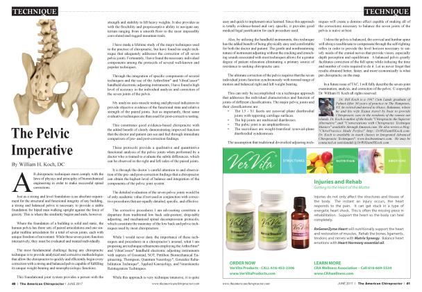
The pelvis is the weight-bearing foundation of the body. It consists of the following seven joints: the symphysis pubis and three paired joints, the 5L-S1 facets, sacroiliacs, and iliofemorals. Each joint must be functionally evaluated for strength and the paired joints for bilateral balance. Because the paired joints and the symphysis pubis are all anatomically and functionally different, they must be corrected accordingly.
The individual techniques that I employ in my examination and corrective protocols were not developed by me. The manner in which I organize them in my protocols is my own work, though. I salute the following doctors who have provided the chiropractic profession with the advanced techniques I employ in my protocols: Dr. George Gonzalez, Quantum Neurology nervous system rehabilitation; Dr. Major DeJamette, sacro occipital technique; Dr. Alan Creed, neural kinetic integration technique; Dr. George Goodheart, applied kinesiology, as taught by Dr. Eugene Charles; Dr. Clarence Gonstead, Gonstead technique; and Dr. Burl Pettibon, spinal biomechanical engineering.
Chiropractic is nothing if it is not specific. There must be an objective indicator for every corrective procedure we perform. The science and art of twenty-first century chiropractic provides us with the ability to give our patients a very high level of evidence-based care.
Part one of this series detailed the examination of the seven pelvic joints. This second part details the corrective procedures. Corrections are only made to address the positive findings of the examination. This allows the body to focus its response only where it is needed. Indiscriminate adjustments dissipate energy. Unnecessary adjustments are usually rejected or cause unpredictable results.
I urge the reader to follow the sequence of corrections I recommend. Each step sets the stage for those that follow until pelvic balance has been achieved. I prefer to complete my entire examination before I begin correction in order to analyze the complete picture of the patient’s presentation.
Correction Procedures
Psoas: I begin with a bilateral psoas release. This eliminates a major source of resistance to the correction.
Although one is likely to be more contracted than the other, I release both because both are predictably hypertonic due to the excessive time most people spend sitting. (See my October 2017 The American Chiropractor article on the psoas.)
Procedure: Patient supine. For the right psoas, the doctor stands on the left side of the patient. With the right leg drawn up 90 degrees to the table, contact the right lower abdomen immediately above the pubic arch with the heel of your right hand. Comfortable pressure is applied A - P and I - S while the knee is maneuvered medially to create a comfortable stretch. A similar stretch can also be applied with the knee maneuvered laterally. Do the same on the left side with the doctor standing on the right using his or her left hand. Then recheck the psoas length by extending the arms overhead palms together with fingers extended and comparing arm lengths. They should now be even.
Note the following contraindications for this procedure: confirmed or suspected pregnancy, excessive abdominal pain or rebound tenderness, or suspicion of an abdominal aortic aneurysm. To check for the possibility of an aneurysm, place your hand on the abdomen and note any strong or broad-based pulse.
Symphysis Pubis: Because of the intimate and sensitive nature of this area, care must be taken to explain the procedure and show on a model spine what you will be doing and why it is important. Secure the patient’s permission before you touch the pubic arch. The doctor’s professional attitude and demeanor are most important and circumvents any misunderstanding.
Procedure: With the patient supine, bring the knees up with feet on the table. Instruct the patient to hold a roll of paper towels or a foam roll between the knees. Place the wide padded bifurcated tip of the ArthroStim adjusting instrument on the superior aspect of the center of the pubic arch. Instruct the patient to squeeze the roll between their knees as the doctor delivers six to 12 light impacts S - I and slightly A - P. Do this three to four times and then retest the ability of the patient to strongly adduct the knees. Increased ability is indicative of correction.
Femur Heads: Femur heads are examined left and right both anteriorly and posteriorly. Correction is not an osseous adjustment, but it is designed to reset the mechanoreceptors located in the joint capsules.
Procedure: For a femur head that tests weak on anterior, the patient is supine with the knee on the weak side raised to 90 degrees. Using the narrow bifurcated tip of the ArthroStim, apply light impact forces around the femur
head as the knee is repeatedly extended and then returned to 90 degrees. Firm resistance on retesting the hip flexion indicates successful correction.
For a femur head that tests weak on posterior, the patient is prone with the knee of the weak side bent to 90 degrees. Instruct the patient to lift the leg off the table (with doctor’s assistance). Using the small bifurcated tip of the ArthroStim instrument, impact lightly around the periphery of the posterior aspect of acetabulum as the leg is moved up and down. Then impact the lumbar spine L5 through LI while continuing to move the leg with the bent knee up and down. Finally, impact the ischium while continuing to move the leg.
The goal is to reset the mechanoreceptors on the anterior and posterior of the femur heads and to restore freedom of motion in the acetabulum. Weakness may be found on left, right, or both hips and anteriorly, posteriorly, or both. Correction is complete when retesting indicates all are strengthened.
The 5L — SI Facets
Next, return the patient to supine and retest the facets. In many cases, resetting the posterior femur head mechanoreceptors coincidentally strengthens 5L - SI facets that had previously tested weak. If the facet still tests weak, a direct decompression facet adjustment is indicated.
Procedure: With the patient supine, raise the leg straight up 30 degrees and lateral 30 degrees. Grasp the calcaneus with one hand and the superior of the foot below the subtalar joint with the other. Give several quick, strong S to I pulls. This will decompress imbricated or jammed facets. An audible release is often heard and felt at the ankle joint. This represents the added benefit of resetting old, preexisting ankle sprains. A formerly weak facet muscle test should test strong after this procedure. If not, repeat decompression.
Sacroiliac or Lumbosacral Joint
The last correction is for either a sacroiliac or lumbosacral subluxation. A weak standing muscle test with positive SOT category two indicators is indicative of a sacroiliac slippage and separation. A weak seated muscle test in the absence of category two indicators suggests a primary lumbosacral subluxation. See part one of this series for details on how to test.
In the majority of cases with lower back pain, especially during the early phase of care, you will find a positive category two sacroiliac slippage and separation. This is designated PRSS or PLSS, according to the short leg side.
Procedure: For category-two treatment, the patient is supine, the short leg side SOT block is inserted under the iliac crest perpendicular to the spine. The long leg block is inserted under the acetabulum and directed 45 degrees superior toward the opposite block. Weak arm fossa tests should be monitored until they strengthen, at which point the blocks are removed and correction is complete.
The patient is then instructed to get off the table and walk around the room. The standing muscle test is repeated. Successful pelvic correction is confirmed when an originally weak standing muscle test is strengthened.
For a primary lumbosacral subluxation, correction may be approached in a number of ways. I usually begin with the patient seated and make impact with the wide bifurcated tip on the ArthroStim instrument as I move it along the lumbar spine. I instruct the patient to move into extension on inspiration and flexion on expiration as I continue to impact with the instrument along the lumbars.
I follow the instrument work with either an SOT category three blocking procedure or a Gonstead side posture lumbosacral adjustment using an AS listing on the long leg side and a PI listing on the short leg side.
The category three correction is performed with the patient prone. The short leg block is inserted under the acetabulum and directed inferiorly 45 degrees. The long leg block is inserted under the ASIS and directed 45 degrees inferiorly.
Successful pelvic correction is confirmed when originally weak standing or seated muscle tests become strong. If a strong test is weakened by cervical flexion, it is indicative of a cranial/cervical subluxation that still needs correction. Cervical and cranial examination and correction will be the subject of a future article.
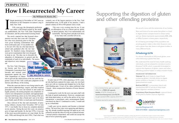
Dr. William H. Koch, a 1967 Cum Laude graduate of Palmer College, practiced in The Hamptons, NY for 30 years and in The Bahamas for 15 years aboard his motor yacht, The Coastal Chiropractor. He currently splits his time between Abaco, Bahamas and his newest practice in Mount Dora, Florida. Dr. Koch is author of the books “Chiropractic the Superior Alternative” and Conversations with Chiropractic Technique Masters” available through Amazon, com. He writes a blog: “ChiroPractice Made Perfect ” http:// DrWilliamHKoch. com and teaches classes on The Koch Protocols for Integrated Advanced Chiropractic Techniques. He may be contacted via email at outislanddc a drwilliamhkoch. com. Learn more by visiting http://KochSeminars.com.