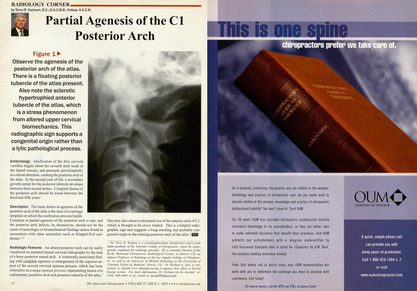
Embrylology. Ossification of the first cervical vertebra begins about the seventh fetal week at the lateral masses, and proceeds perichondrally in a dorsal direction, creating the posterior arch of the atlas. In the second year of life, a secondary growth center for the posterior tubercle develops between these neural arches. Complete fusion of the posterior arch should be noted between the third and fifth years.1 Description. The basic defect in agenesis of the posterior arch of the atlas is the lack of a cartilage template on which the ossification process builds. Complete or partial agenesis of the posterior arch is rare, and the posterior arch defects, by themselves, should not be the cause of neurologic or biomechanical findings unless found in association with other anomalies such as Klippel-Feil syndrome. 1-u Radiologic Features. An absent posterior arch can be easily visualized on standard lateral cervical radiographs by the lack of a bony posterior neural arch. A commonly associated finding with complete agenesis is enlargement of the superior aspect of the second cervical spinous process, which has been referred to as a mega-spinous process, representing fusion of a rudimentary posterior arch and posterior tubercle of the atlas.2 One may also observe increased size of the anterior arch of C1, which is thought to be stress related. This is a helpful radio-graphic sign and suggests a long-standing and probable congenital origin to the missing posterior arch of the atlas.' | Dr. Terry R. Yochum is a second-generation chiropractor and a cum laude graduate of the National College of Chiropractic, where he subsequently completed his radiology specialty. He is currently Director of the Rocky Mountain Chiropractic Radiological Center, in Denver, CO, an Adjunct Professor of Radiology at the Los Angeles College of Chiropractic, as well as an instructor of Skeletal Radiology at the University of Colorado School of Medicine, Denver, CO. Dr. Yochum is, also, a consultant to Health Care Manufacturing Company that offers a Stored Energy system. For more information. Dr. Yochum can be reached at: (303J 940-9400 or by e-mail at dcrad099(a)anl.com. Figure 1 Observe the agenesis of the posterior arch of the atlas. There is a floating posterior tubercle of the atlas present. Also note the sclerotic hypertrophied anterior tubercle of the atlas, which is a stress phenomenon from altered upper cervical biomechanics. This radiographic sign supports a congenital origin rather than a lytic pathological process.