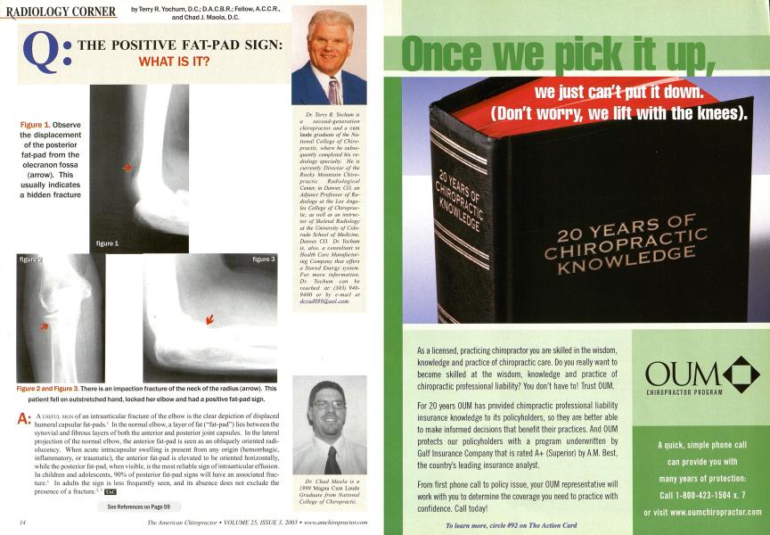
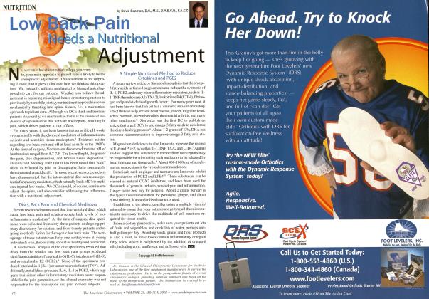
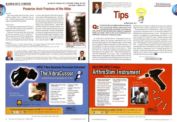
A useful sign of an intraarticular fracture of the elbow is the clear depiction of displaced humeral capsular fat-pads.' In the normal elbow, a layer of fat ("fat-pad") lies between the synovial and fibrous layers of both the anterior and posterior joint capsules. In the lateral projection of the normal elbow, the anterior fat-pad is seen as an obliquely oriented radi-olucency. When acute intracapsular swelling is present from any origin (hemorrhagic, inflammatory, or traumatic), the anterior fat-pad is elevated to be oriented horizontally, while the posterior fat-pad, when visible, is the most reliable sign of intraarticular effusion. In children and adolescents, 90% of posterior fat-pad signs will have an associated fracture.1 In adults the sign is less frequently seen, and its absence does not exclude the presence of a fracture." •' BW3 See References on Page 59 Dr. Terry R. Yochum is a second-generation chiropractor and a cum laude graduate of the National College of Chiropractic, where he subsequently completed his radiology specialty. He is currently Director of the Rocky Mountain Chiropractic Radiological Center, in Denver, CO, an Adjunct Professor of Radiology at the Los Angeles College of Chiropractic, as well as an instructor of Skeletal Radiology at the University of Colorado School of Medicine, Denver, CO. Dr. Yochum is, also, a consultant to Health Care Manufacturing Company that offers a Stored Energy system. For more information, Dr. Yochum can be leached at: (S03) 940-9400 or by e-mail at [email protected] Dr. Chad Maola is a 1999 Magna Cum Laude Graduate from National College of Chiropractic. Figure 1. Observe the displacement of the posterior fat-pad from the olecranon fossa (arrow). This usually indicates a hidden fracture Figure 2 and Figure 3. There is an impaction fracture of the neck of the radius (arrow). This patient fell on outstretched hand, locked her elbow and had a positive fat-pad sign.