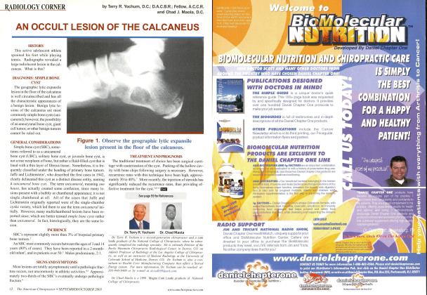
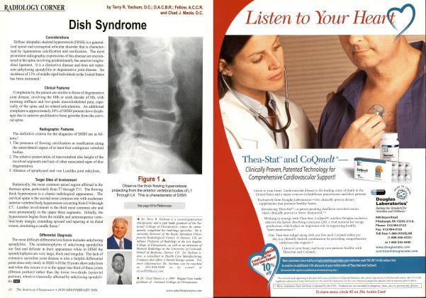
CASE HISTORY This young adult patient presents with neck pain following an MVA. You note an abnormality in the upper cervical spine atCl & C'2. Is this a fracture? DISCUSSION Anomalies ol" the odontoid process are considered uncommon (1) and are usually discovered by the principle "traumatic determinism." This means that the underlying condition predated the injury and is not caused by this current trauma. These anomalies may be associated with Down's syndrome, Klippel-Feil syndrome, Morquio's syndrome, and spondyloepiphy-seal dysplasia. Clinical Features: Any symptoms the patient may manifest are usually the result of atlantoaxial instability with resultant cord compression; however, if there is compression of the vertebral artery resulting from stretching of the artery during Cl subluxation on C2, then the symptoms may be considerably greater. Increased deep-tendon reflexes, proprioceptive loss, or sphincter incompetence may be encountered. Additionally, compression of the vertebral arteries may result in local thrombosis and vascular occlusion. The thrombus may also serve as a source for emboli to the brain. Clearly, the combination of os odontoideum with high-velocity injury can produce central cord syndrome or even fatal injury. Radiologic Features: The X-ray diagnosis of os odontoideum in a child below the age of 5 years can be made, if there is demonstration of hy-permobility of the odontoid process on the body of C2 during flexion and/or extension. In the adult, an X-ray diagnosis is certain, if a smooth, wide, lucent defect is seen to separate the odontoid process from the C2 body at the level of the superior articular processes, and there is an associated stress hypertrophy (enlargement) of the anterior tubercle of the atlas. This finding will not be present in the child, as the biomechanical stresses on the anterior arch of the atlas will not have been present for a long enough period to allow the hypertrophy to develop. Os odontoideum must be differentiated from an acute fracture of the odontoid process. A helpful radiographic sign that may be present and that confirms a developmental defect of the odon- toid process is a "molding: of the anterior arch of Cl into the ventral aspect of the odontoid process. Magnetic resonance imaging is useful in evaluating the spinal cord for angulation, compression, and intramedullary injury (contusion). Dr. Terry R. Yocliuni is a second generation chiropractor and a Cum Laude Graduate of National College of Chiropractic, where he subsequently completed his radiology residency. He is currently Director of the Rocky Mountain Chiropractic Ra- diological Center in Denver, CO, and Adjunct Professor of Radiology at the Southern California University of Health Sciences, as well as an instructor of skeletal radiology at the University of Colorado School of Medicine, Denver, CO. Dr. Yochum can he reached at 1-303-940-9400 or hy e-mail at dcrad()99(aaol.com. Dr. Chad Maola is a 1990 Magnci Cum Laudc Graduate of National College of Chiropractic. Dr. Maola is available for post-graduate seminars. He may he reached at 1-727-433-0153 or bv email at de Ol(a msn.com. Reference 1 .Yochum. T. R.. Rowc. L.J.: Essentials of Skeletal Radiology, 3rd ed., Williams & Wilkins, Baltimore, Maryland, 2005.BH Figure 1 Note the odontoid is not attached to the body of C2 and the C1 anterior tubercle is enlarged from stress hypertrophy. Figure 2. Tomogram Note the smooth margin to the base of the odontoid indicating no fracture and an os odontoideum.