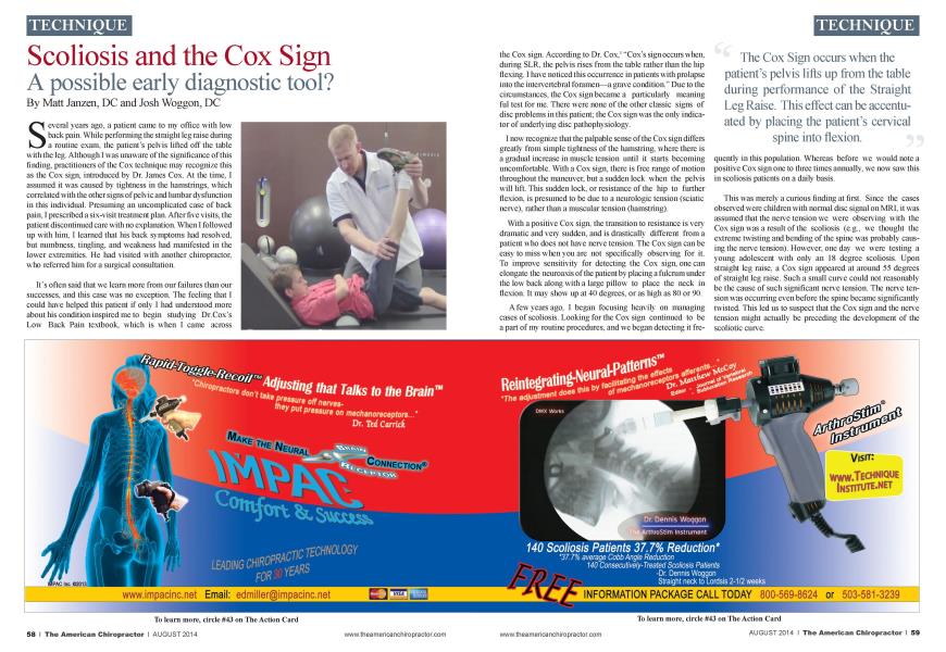
S everal years ago. a patient came to my office with low back pain. While performing the straight leg raise during a routine exam, the patient's pelvis lifted off the table with the leg. Although I was unaware of the significance of this finding, practitioners of the Cox technique may recognize this as the Cox siga introduced by Dr. James Cox. At the time. I assumed it was caused by tightness in the hamstrings, which correlated with the other signs of pelvic and lumbar dysfunction in this individual. Presuming an uncomplicated case of back pain. I prescribed a six-visit treatment plan. After five visits, the patient discontinued care with no explanation. When I followed up with him. I learned tliat his back symptoms had resolved, but numbness, tingling, and weakness hid manifested in the lower extremities. He had visited with another chiropractor, who referred him for a surgical consultation. It's often said that we learn more from our failures than our successes, and this case was no exception. The feeling that I could have helped this patient if only I had understood more about his condition inspired me to begin studying Dr.Cox"s Low Back Pain textbook, which is when I came across the Cox sign. According to Dr. Cox.1 "Cox"s sign occurs when, during SLR. the pelvis rises from the table rather than the hip flexing. I have noticed this occurrence in patients with prolapse into the inten crtebral foramen—a grave condition." Due to the circumstances, the Cox sign became a particularly meaning ful test for me. There were none of the other classic signs of disc problems in this patient; the Cox sign was the only indicator of underlying disc pathophysiology. I now recognize that the palpable sense of the Cox sign differs greatly from simple tightness of the hamstring, where there is a gradual increase in muscle tension until it starts becoming uncomfortable. With a Cox sign, there is free range of motion throughout the maneuver, but a sudden lock when the pelvis will lift. This sudden lock, or resistance of the hip to further flexion, is presumed to be due to a neurologic tension (sciatic nerve), rather than a muscular tension (hamstring). With a positive Cox sign, the transition to resistance is very dramatic and very sudden, and is drastically different from a patient who does not have nerve tension. The Cox sign can be easy to miss when you are not specifically observing for it. To improve sensitivity for detecting the Cox sign, one can elongate the neuroaxis of the patient by placing a fulcrum under the low back along with a large pillow to place the neck in flexion. It may show up at 40 degrees, or as high as 80 or 90. A few years ago. I began focusing heavily on managing cases of scoliosis. Looking for the Cox sign continued to be a part of my routine procedures, and we began detecting it fre- qucntly in this population. Whereas before we would note a positive Cox sign one to three times annually, we now saw this in scoliosis patients on a daily basis. This was merely a curious finding at first. Since the cases observed were children with normal disc signal on MRI. it was assumed that the nerve tension we were observing with the Cox sign was a result of the scoliosis (e.g.. we thought the extreme twisting and bending of the spine was probably causing the nerve tension). However, one day we were testing a young adolescent with only an 18 degree scoliosis. Upon straight leg raise, a Cox sign appeared at around 55 degrees of straight leg raise. Such a small curve could not reasonably be the cause of such significant nerve tension. The nerve tension was occurring even before the spine became significantly twisted. This led us to suspect that the Cox sign and the nerve tension might actually be preceding the development of the scoliotic curve. In 1968. Dr. Roth first proposed the concept of nerve tension, specifically tension in the spinal cord, as the cause of scoliosis. Dr. Roth proposed that a spinal cord that was short relative to the length of the vertebral column might result in a tension that causes the spine to "coil down" around the axis of the spinal cord. Such a theory provides a logical explanation for the paradoxical rotation of the vertebra in a scoliosis. in that the vertebra rotate opposite to the spinal cord." Dubousset observed that, in any type of spine, the spinal cord will always follow the shortest route through the canal, presumabh to reduce the tension on the nerves as much as possible.' It wasn't until decades later that more researchers began referencing Dr. Roths theory. The theory has been termed "uncoupled ncuro-osseous development." which, according to Porter, states that "in some patients with scoliosis there is disproportionate neuro-osscous growth—the longitudinal growth of the spinal cord fails to keep pace with the growth of the vertebral column and. as a consequence, the spine buckles into a scoliosis deformity."5 Porter points out that many abnormalities associated with scoliosis can be explained well, in that they may contribute to asynchronous neuro-osseous development and. hence, spinal cord tension. For example, dysfunction of the pineal gland and melatonin has been implicated in scoliosis. Such dysfunction of the endocrine system is theorized to contribute to an asynchronous neuro-osscous development. Since Porter's article in 2001. there has been a growing awareness of this theory in the published literature.67 Dr. Cox proposed the Cox sign as an observation of extreme nerve tension caused by more severely entrapped nerve roots. Now we are seeing tliat same Cox sign in a child-onset condition in which nerve tension is the leading theory for causation. If the Cox sign is truly and reliably indicative of underlying nerve tension in a child, it may help to identify individuals who are at risk for developing scoliosis. My personal story supporting this theory occurred with the appearance of the Cox sign in my daughter when she was six and a half years old. I was shocked the evening I first observed this, realizing the potential implication for my own daughter. I followed up with a postural evaluation and scoliometry . yet there was no evidence of any curvature or rotation as would occur in scoliosis. However, if the Cox sign was indicative of a nerve tension tliat can cause scoliosis. it meant that my daughter was at very high risk fora future scoliotic curve. Over the course of a year, the Cox sign persisted. When she was eight years old. we performed a standing 3-D full-spine MRI. which revealed a 13 degree left lumbar scoliosis. When seated, the MRI revealed a 21 degree scoliosis—a common finding of a true scoliosis for the curve size to increase as the patient sits versus stands. It must be recognized that idiopathic scoliosis (IS) is not a diagnosis, but rather a lack of one. Multiple, distinct, and separate disease entities will fall under the umbrella term of IS cases tested in our office, obyious clinical signs of nerve tension are consistently found. Currently, difficult and time-consuming MRI techniques are being researched to uncover eyidence of spiral cord tension in IS cases. Expensive and complex genetics tests are being developed to attempt to predict risk for progression. All of this technology still only catches the problem quite late in the game—only after a scoliosis has had a chance to significantly deform the structure of a spine. If the Cox sign proves to be a reliable predictor of a root-driving force for IS. then detecting scoliosis before it occurs may actually become a reality. If the Cox Test could prove to be a quick, inexpensive early detection test for scoliosis. chiropractors and pediatricians could easily perform it routinely to detect scoliosis before it even occurs. While further research is needed to draw conclusions, the chiropractic profession has the opportunity to lead the way in this potentially groundbreaking early scoliosis detection research. Dr. Matt Janzen with more than 17 years of experience in chiropractic, specializing in scoliosis. is a practitioner and received his doctorate degrees through Palmer University as a Salutatorian in 1996. specializes in musculoskeletal condi- lions of the spine and extremities, gearing towards sports-related injuries. This later allowed him to become a Certified Chiropractic Extremity Practitioner (C.C.EP) with the study and practice of Active Release Techniques (ART), which are highly effective for sports injuries. References: Cox, JM: Low Back Pain, Mechanism, Diagnosis, Treatment, 6th Edition. Philadelphia: Lippincott, Williams and Wilkins 1999: pgs 438-439. Roth A /: ldiopathic scoliosis caused by a short spinal coni. Ada RadiolDiagn. 1968 .\(ay;7(3):257-71. HilalSK, Keim HA: Selective spinalangiographv in adolescent scoliosis. Radiology 1972,102:349 359. Chu JVC et al: Relative shortening and functional tethering of spinal con! in adolescent idiopathic scoliosis? Study with mulliplanar reformat magnetic resonance imaging and somatosensoiy evoked potential. Spine. 2006;31 :E19-E25. Porter RW: Can a short spinal cord produce scoliosis? Eur Spine J.Feb2001;10(l):2-9. Chu el a I: Relative shortening and functional tethering of spinal cord in adolescent scoliosis Result ofasynchmnous neuro-osseous growth, summaty of an electmnic focus group debate of the IBSE. Scoliosis 2008:3:8. Burwell etal: Pathogenesis of adolescent idiopathic scoliosis in girls - a double neuro-osseous theory involving disharmony between two nervous systems, somatic and autonomic expressed in the spine and trunk: possible dependency on sympathetic nervous system and hormones with implications for medical therapy. Scoliosis 2009,4:24. Dr. A. Joshua Woggon, a 2010 Graduate of Parker College, serves as the Director of Research for the CLEAR Scoliosis Institute, a Xon-Profit Organization dedicated to advancing chiropractic scoliosis correction (www.clear- institute.org). He can be contacted at [email protected].