Concussion: Diagnosis-Testing-Chiropractic
“New testing is available using biomarkers”
IDENTIFYING
CONCUSSION
Mark Studin
William J. Owens
DC, FASBE(C), DAAPM, DAAMLP
DC, DAAMLP
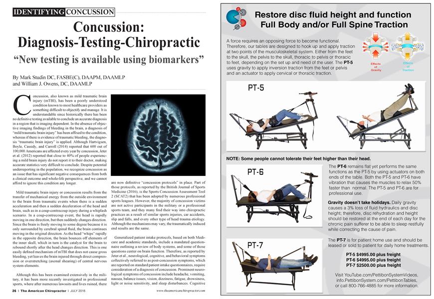
Concussion, also known as mild traumatic brain injury (mTBI), has been a poorly understood condition known to most healthcare providers as something difficult to objectify and manage. It is understandable since historically there has been no definitive testing available to conclude an accurate diagnosis in a region that is imaging dependent. In the absence of objective imaging findings of bleeding in the brain, a diagnosis of “mild traumatic brain injury” has been affixed to the condition, whereas if there is evidence of traumatic bleeding, the diagnosis “traumatic brain injury” is applied. Although Hartvigsen, Boyle, Cassidy, and Carroll (2014) reported that 600 out of 100,000 Americans ai e affected every year by concussion, Jeter et al. (2012) reported that close to 40% of people experiencing a mild brain injury do not report it to their doctor, making accurate statistics very difficult to conclude. Despite potential underreporting in the population, we recognize concussion as an issue that has significant negative consequences from both a clinical outcome and whole-life perspective, and we cannot afford to ignore this condition any longer.
Mild traumatic brain injury or concussion results from the transfer of mechanical energy from the outside environment to the brain from traumatic events when there is a sudden acceleration and then a sudden deceleration of the head and brain, such as in a coup-contrecoup injury during a whiplash scenario. In a coup-contrecoup event, the head is rapidly moving in one direction, but then suddenly changes direction. Since the brain is freely moving to some degree because it is only surrounded by cerebral spinal fluid, the brain continues moving in the original direction. As the head “whips” rapidly in the opposite direction, the brain bounces off elements of the inner skull, which in turn is the catalyst for the brain to rebound shortly after the head changes direction. This is one easily defined mechanism of mTBI that does not cause gross bleeding, yet leaves the brain injured through direct compression or overstretching (axonal shearing) of central nervous system elements.
Although this has been examined extensively in the military, it has been more recently investigated in professional sports, where after numerous lawsuits and lives ruined, there
are now definitive “concussion protocols” in place. Part of those protocols, as reported by the British Journal of Sports Medicine (2016), is the Sports Concussion Assessment Tool 2 (SCAT2) that has been adopted by numerous professional sports leagues. However, the majority of concussion victims are not active participants in the military or a professional sports team, and they many find their way into chiropractic practices as a result of similar sports injuries, car accidents, slip and falls, and every other type of head trauma etiology. Although the mechanisms may vary, the traumatically induced end results are the same.
Generalized patient intake protocols, based on both Medicare and academic standards, include a mandated questionnaire outlining a review of body systems, and some of those questions center on brain function. Therefore, as reported by Jeter et al., neurological, cognitive, and behavioral symptoms collectively referred to as post-concussion symptoms, which are reported on standard patient intake questionnaires, require consideration of a diagnosis of concussion. Prominent neurological symptoms of concussion include headache, vomiting, nausea, balance issues, vision, dizziness, fatigue, drowsiness, light or noise sensitivity, and sleep disturbances. Cognitive
symptoms include deficits in attention, concentration, memory, mental processing speed, and working memory or decisionmaking. Common behavioral symptoms include anxiety, depression, irritability, aggression, and depression. The researchers went on to report that these symptoms can persist in approximately 25% of these cases.
As a profession, chiropractic is a critical part of
the rehabilitation for the concussion population because the post-traumatic patient typically presents to the average chiropractic practice. As chiropractors (along with all healthcare providers), if you combine the history with the previously mentioned symptoms inclusive of neurological, cognitive, and behavioral traits, you then have the direction or “triage road map” of how to conclusively differentially diagnose your patient, including what tests to consider conducting in order to do so. The first line of testing is to consider advanced imaging to rule out bleeding and ensure the patient does not need an immediate neurosurgical consultation. With this set of signs and symptoms, treating blindly can put your patient at possible extreme risk.
■ "More recently, diffusion tensor imaging (DTI) has been a tool available to image mTBI victims that uses tissue water diffusion rates to determine bleeding at a very small level, giving demonstrable evidence to brain injury. J J
Imaging of the brain necessitates either MRI or CAT scans. MRI is more sensitive, and in the absence of bleeding, the diagnosis is limited to mTBI or concussion (used interchangeably). More recently, diffusion tensor imaging (DTI) has been a tool available to image mTBI victims that uses tissue water diffusion rates to determine bleeding at a very small level, giving demonstrable evidence to brain injury. As reported by Soares, Marques, Alves, and Sousa (2013), DTI has multiple issues to overcome in order to certify accuracy, including but not limited to tissue type, integrity, barriers, and quantitative diffusion rates that aie required to infer molecular diffusion rates. Currently, DTI is a model based upon assumption with a very promising outlook as a reliable tool.
Historically, mTBI was exclusively diagnosed by an omission of advanced imaging findings and the presence and persistence of the neurological, cognitive, and behavioral signs and symptoms. Today, brain-derived neurotrophic factors (BDNF) offer answers about post-traumatic brain pathology that are both conclusive and reproducible. According to Korley et al.
(2015), BDNF is a secreted neurotrophin (chemical hormone or messenger in blood) that promotes the development, maintenance, survival, differentiation, and regeneration of neurons. BDNF also is important for synaptic plasticity (strengthening of synapses over time) and memory processing. Germane to mTBI and concussion, BDNF has been implicated in reducing secondary brain injury, with elevations providing neuroprotection and restoring connectivity traumatic brain injury.
Korley et al. went on to report that BDNF levels were the highest in the normal group with lower values in mTBI and even lower in traumatic brain injury (TBI) subjects. In addition, very low BDNF values were more often associated with incomplete recovery of mTBI patients than moderate or severe TBI patients. As a result, it has been determined that BDNF has a higher prognostic value for identifying mTBI related sequelae at six months.
Korley et al. continued to note that BDNF is the most abundantly secreted brain neurotrophin and a secreted protein, and it can be readily measured using well-established immuneassay techniques, identifying it as a non-necrosis brain injury biomarker. This distinguishes BDNF from other protein-based biomarkers that are structural components of neurons and myelin-based proteins among other neurologic structures. In order for structural proteins to be found in high abundance in circulation, sufficient cellular necrosis and damage to the blood barrier membrane must be observed. However, BDNF
does not require cellular damage or necrosis to be observed in circulation, allowing BDNF to be more abundant in circulation than structural proteins.
After a traumatic brain event, BDNF supports synaptic reorganization and restoration during the brain circuitry “reconnection” phase. Therefore, lowered BDNF values indicate a better prognosis. In patients with a BDNF comorbidity of anxiety, depressive disorders, and schizophrenia, low BDNF values on the day of the injury predispose this population to incomplete recovery as a risk factor. Korley et al. concluded that serum BDNF discriminates between mTBI and TBI cases with excellent diagnostic accuracy. Additionally, lowered BDNF values are associated with incomplete recovery and are useful in identifying patients that are likely to retain symptoms six months after trauma.
Simply put, a blood test could assist providers in concluding the presence and/or severity of traumatic brain injury or mild traumatic brain injury. The results afford an early diagnosis so that you can devise a treatment plan inclusive of altering activities of daily living to prevent further damage and maximize the repair process with minimizing further physical, chemical, or emotional stressors.
Based upon interviews with leading neurologists and neurosurgeons who understand and have first-hand experience of both receiving chiropractic care and managing and treating
mTBI patients, it is recommended that, until the signs and symptoms of the neurological, cognitive, and behavioral abate, high-velocity rotational cervical adjustments should be avoided to allow the brain to “repair and rewire” the connections without further possibilities of coupcontrecoup energy to the brain. We concur with this recommendation while recognizing that chiropractic care should not be avoided, just adapted to allow the brain to heal.
References:
1. Hartvigsen,./., Boyle, E., Cassidy,./. D., & Carroll, L../. (2014). Mild traumatic brain injury after motor vehicle collision: What are the symptoms and who treats them? A population-based 1-year inception cohort study. Archives of Physical Medicine and Rehabilitation, 95(Suppl. 3), S286-S294.
2. Jeter, C. B., Hergenroeder, G. W, Hylin, M../.. Redell, J. B., Moore, A. N, & Dash, P. K. (2013). Biomarkers for the diagnosis and prognosis of mild traumatic brain injury/concussion. Journal of Neurotrauma, 30(8), 657-670.
3. British Journal of Sports Medicine. (2016). Sport concussion assessment tool 2. Retrieved from http://bjsm.bmj.com/content/43 Suppl 1/Í85.full.pdf
4. Soares, J. M., Marques, P, Alves, V, & Sousa, N. (2013). A hitchhiker ’s guide to diffusion tensor imaging. Frontiers in Neuroscience, 7(31), 1-14.
5. Korley, F. K., Diaz-Arrastia, R., Wu, A. H. B., Yue, J. K., Manley, G. T, Sair, H. I., VanEyk,./., Everett, A. I)., Okonkwo, D. O., Valadka, A. B., Gordon, W. A., Maas, A. I., Mukherjee, P, Yuh, E. L., Lingsma, H. F, Puccio, A. M., & Schnyer, D. M., (2015). Circulating brain-derived neurotrophic factor has diagnostic and prognostic value in traumatic brain injury. Journal of Neurotrauma, 32,1-11.
^^SZ Dr. Mark Studin is an adjunct associate professor of chiropractic at the University of Bridgeport Col^ C 'M lege of Chiropractic; an adjunct professor of clinical BfC Æ sciences at Texas Chiropractic College; and a clinical I presenter for the State of New York at Buffalo, School l àÂH of Medicine and Biomedical Sciences for postdoctoral education, teaching MRI spine interpretation, spinal biomechanical engineering, and triaging trauma cases. He is also the president of the Academy of Chiropractic teaching doctors of chiropractic how to interface with the medical and legal communities (www. DoctorsPIProgram.com); teaches MRI interpretation and triaging trauma cases to doctors of all disciplines nationally; and studies trends in health care on a national scale (www. TeachDoctors.com). He can be reached at DrMark(xi)AcademyofChiropractic.com or at 631-786-4253.
Dr Bill Owens is presently in private practice in Buffalo and Rochester, New York, and generates the majority of his new patient referrals directly from the primary care medical community. He is an associate adjunct professor at the State University of New York at Buffalo School of Medicine and Biomedical Sciences and at the University of Bridgeport, College of Chiropractic, and an adjunct professor of clinical sciences at Texas Chiropractic College. He also works directly with doctors of chiropractic to help them build relationships with medical providers in their community. He can be reached by e-mail at dr owensdecade my of chiropractic, com, through www.mdreferralprogram.com, or by calling 716-228-3847.