Orthotics for Women
ORTHOTICS
John K. Hyland
DC, DACBR, DABCO, CSCS
Women and men are different in structure and biomechanics. This statement, hopefully, doesn’t require a footnote to support its accuracy. Women’s feet are just like the rest of their musculoskeletal system, but they are, in fact, among the most different of the structures. In spite of this, we often tend to treat female patients who have foot imbalance and lower-extremity dysfunction the same way that we treat male patients. It is vitally important to remember and review many important areas of difference so that we can provide specific and effective care.
Women’s feet are different in shape and size. They are encouraged to wear very different shoes and their knee alignment and gait styles are very dissimilar. Joint problems and symptom patterns differ, so orthotic solutions need to be specific. As women age, their foot problems tend to become more severe, often resulting in significant disability and problems with walking.1 Appropriate lower-extremity treatment and spinal support depend on differentiating each woman’s needs.
Women ’s Feet Foot Shape
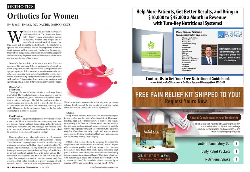
The shape of a woman’s foot varies in several ways from a man’s foot. The female foot tends to have a narrower heel (in relation to the forefoot), and is narrower overall than a man’s foot, relative to its length.2 The Achilles tendon is smaller in circumference and strength, but it is also shorter. Because of the narrow heel and foot, the forefoot is relatively quite wide. This causes the biomechanical forces on the foot to be distributed in a different manner.
Foot Problems
Women tend to develop biomechanical problems and symptomatic conditions in the forefoot more frequently than men do. Bunions (hallux valgus), hammertoes, callus formation, interdigital neuromas, and metatarsalgia are all more common in women.3 Many of these conditions have been linked to abnormal biomechanical forces in the feet.
Using weight-bearing radiographs, researchers demonstrated the correlation of hallux valgus and medial arch collapse. They found that “the single most dominant variable affecting metatarsal pronation (and hallux valgus) was the height of the medial longitudinal arch.”4 Using a different approach, other investigators compared weight-bearing X-rays of the hindfeet in normal female subjects to those with hallux valgus. They found that the calcaneus and talus in the feet with hallux valgus had excessive pronation.5 Another recent study has confirmed that callus formation is closely associated with several specific “abnormal foot weight-bearing patterns.”
These patterns are a lower medial arch with greater pronation; reduced dorsiflexion of the first metatarsal joint; and limited ankle dorsiflexion (due to calf muscle tightness).6
Solution
Every woman needs to wear shoes that have been designed for the gender-specific needs of the female foot. This means that they need a shoe that is narrow at the heel and widens substantially at the forefoot. It should have a short but wide toe box and some mechanism (such as laces or straps) to keep the narrow heel in place during gait. Unfortunately, this describes very few of the shoes currently bought and worn by women in our society. Certainly, traditional high heels and pumps do not fall into the healthy shoe category.
Orthotics for women should be designed to support the longitudinal and anterior transverse arches,7 as well as provide metatarsal padding and limit excessive heel motion. Researchers have found that small, dense metatarsal arch pads positioned more distally are most effective in reducing the pressures on the metatarsal heads.8 One study of patients with metatarsalgia found that custom-made orthotics with a “metatarsal dome” decreased the plantar pressures by an average of 17% and reduced the reported pain by 71%.9
Women ’s Shoes Fashion Victims
The fashion industry perpetuates the image of the sexy woman in high-heeled shoes. Many women wear heels that plantarflex the ankle and place increased pressures on the forefoot and metatarsal heads. Even a heel as low as threefourths of an inch has been found to increase the pressure on the forefoot by as much as 22%.10
Sizes and Fit
Constrictive shoes accentuate the problems of high heels, and they create problems of their own. A study of 356 healthy women found that 88% were wearing shoes that were significantly smaller than their feet.11 The average difference measured between the shoe and the foot was 1.2 cm! Eighty percent of the women in the study stated that they had some foot pain (almost all in the forefoot). Interestingly, for those women who reported no foot pain, the average foot-shoe discrepancy was only 0.56 cm. The researchers recommended that women should always buy their shoes by fit and not by size, and that shoes should always be tried on at the end of the day and be fitted to the weight-bearing foot.
Solution
All female patients should have their footwear checked for fit since many are wearing shoes that don’t fit their feet, and with heel heights that increase the pressures on the fronts of their feet. One easy method to check shoe fit is to trace each foot while standing and then trace the shoe (as was done in the shoe survey previously described). Any significant discrepancy means that the foot is cramped when standing and restricted during gait. When a higher heel is worn, more pressure is exerted on the forefoot, making proper fit more critical.
A properly designed orthotic support should maintain all of the arches, and padding and support for the anterior transverse (metatarsal) arch is especially important for women. However, no orthotic can support the foot correctly if it is placed in an improperly fitted shoe. Shoe size, both length and width, must allow for correct biomechanics during gait. Most women need to have their feet rechecked for correct size, and many will need to purchase new shoes. Orthotics are now available to fit in many different styles of shoes, so proper selection is important. Virtually all women will need more than one pair of orthotics—one pair for work and dress shoes, and another pair (with more shock absorption) designed to fit into recreational shoes.
Women ’s Knees
Patellar Tracking
The Q angle measures the alignment of the quadriceps muscle’s pull from the pelvis to the patella, and the patellar tendon’s pull from the tibia. Since large forces aie transmitted through the patella during knee movement, any increase in the angle will result in tracking problems and eventually symptoms will arise. We know that women tend to have higher Q angles than men. The normal range ends at 15 degrees in males and 20 degrees in females.12 Whenever a patient has excessive pronation of the foot, Q angle stresses are magnified. Prolonged time in pronation causes excessive internal rotation of the tibia, impeding its normal external rotation during gait progression in the stance phase.13 This excessive internal tibial rotation transmits abnormal forces upward in the kinetic chain and produces medial knee stresses, force vector changes of the quadriceps mechanism, and lateral tracking of the patella.14 The combination of a higher Q angle with excessive pronation causes many women to progress from knee dysfunction to patellofemoral arthralgia to degenerative joint disease in the knee.
Anterior Cruciate Ligaments
Female athletes appear to sustain knee injuries more frequently than their male counterparts in similar sports.15 Many of these are noncontact ruptures of an anterior cruciate ligament (ACL).16
Many theories have been proposed to explain the greater
susceptibility of women to ACL tears. Some of these include a smaller intercondylar notch, hormonally mediated ligament laxity, and gender-related differences in muscle strength and balance. One of the most promising concepts considers the alignment of the foot and knee during strenuous sports activities. Researchers have found that whenever the leg is internally rotated, the ACL is much more susceptible to injury. One study by Arnold et al. found that 81% of athletes with injury to the ACL recalled the moment of injury as having their tibia in internal rotation combined with a sudden change of direction at foot strike.17 Other researchers have found excessive pronation of the foot and collapse of the arch during weight bearing to be more common in ACL-injured subjects, and proposed that the subsequent excessive tibial rotation was the mechanism of their injury.18 Another study found that ruptures of the ACL in female athletes were directly correlated with the amount of arch collapse and hyperpronation.19
Solution
The most effective way to decrease a high Q angle and to lower the rotational stress on the knee joint is to prevent excessive pronation with custom-fitted foot orthotics.20 The orthotics should support the foot’s arches (but especially the medial longitudinal arch) and may need to include a pronation wedge under the calcaneus. One study found that using soft corrective orthotics was more effective in reducing patellofemoral pain in women than a physical therapy exercise program.21
Conclusion
All patients should have their footwear checked for fit, but this is especially important for women, who are often wearing poorly fitted shoes and whose higher heels can significantly increase the forefoot pressures. Research has shown that many lower-extremity conditions in women are associated with or directly caused by abnormal biomechanics of their feet. A properly designed orthotic support for women should maintain all of the arches and reduce impact loading. However, padding and support for the anterior transverse (metatarsal) arch, and control of overpronation at the heel appear to be particularly important. Remember that no orthotic can support the foot correctly if it is placed in an improperly fitted shoe. Shoe size, both length and width, must allow for correct biomechanics during gait. For many women, education regarding shoe fit and addressing lower-extremity biomechanics will help prevent some of the disability commonly associated with aging.
References:
1. Leveille SG, Guralnik JM, Ferrucci, L, et al. Foot pain and disability in older women. Am J Epidemiol. 1998;148:657-65.
2. Frey C. Foot health and shoewear in women. Clin Orthop. 2000;372:32-44.
3. Rudicel SA. Evaluating and managing forefoot problems in women. J Musculoskel Med. 1999;16:562-7.
4. Eustace S, et al. Hallux valgus, first metatarsal pronation and collapse of the medial longitudinal arch—a radiological correlation. Skeletal Radiol.1994;23:191-4.
5. Tanaka Y, et al. Hindfoot alignment of hallux valgus evaluated by a weightbearing subtalar x-ray view. Foot Ankle Int. 1999;20:640-5.
6. Bevans JS, Bowker R Foot structure and function: etiological risk factors for callus formation in diabetic and non-diabetic subjects. The Foot. 1999;9:120-7.
7. Chou LB. Disorders of the first metatarsophalangeal joint. Phys Sports.
Med 2000;28.
8. Hayda R, et al. Effect of metatarsal pads and their positioning: a quantitative assessment. Foot & Ankle. 1994;15:561-6.
9. Poon C, Love B. Efficacy of foot orthotics for metatarsalgia. Foot Inf 1. 1997;7:202-4.
10. Snow R. Williams K, Holmes G. The effects of wearing high-heeled shoes on pedal pressure in women. Foot Ankle. 1992;13:85-92.
11. Frey C, et al. American orthopedic foot and ankle society women's shoe survey. Foot Ankle. 1993;14:78-81.
12. Hvid I, Anderson LB, Schmidt H. Chondromalacia patellae: the relation to abnormal patellofemoral joint mechanics. Acta Orthop Scand. 1981;52:661-9.
13. Zappala GG, Taffel CB, Scuderi GR. Rehabilitation of patellofemoral joint disorders. Orthop Clin North Am. 1992;23:555-66.
14. Tiberio D. The effect of excessive subtalar joint pronation on patellofemoral mechanics: a theoretical model. J Ortho Sports Phys Therap. 1987;9:160-5.
15. DeHaven K. Litner K. Athletic injuries: Comparison by age, sport, and gender. Am J Sports Med. 1986;10:218-24.
16. Arendt EA. Common musculoskeletal injuries in women. Phys Sportsmed. 1996;24:39-47.
17. Arnold HA, et al. Natural history of the anterior cruciate ligament. Am J Sports Med. 1979;7:305-13.
18. Beckett ME, et al. Incidence of hyperpronation in the ACL injured knee: a clinical perspective. J Athl Train. 1992;27:58-62.
19. Loudon JK. The relationship between static posture and ACL injury in female athletes. J O S PT. 1996;24:91-97.
20. D'Amico JC, Rubin M. The influence of foot orthotics on the quadriceps angle. JAm Podiatr Med Assoc. 1986;76:337-40.
21. Eng JJ, Pierrynowski MR. Evaluation of soft foot ortho tics in the treatment of patellofemoral pain syndrome. Phys Ther. 1993;73:62-70.
A 1980 graduate of Logan College of Chiropractic, Dr. John Hyland practiced for more than 20 years in Colorado. In addition to his specialty board certifications in chiropractic orthopedics (DABCO) and radiology (DACBR), Dr. Hyland is nationally certified as a strength
and conditioning specialist (CSCS) and a health education specialist
(CHES). He has consulted chiropractors in the concepts andprocedures
of spinal rehabilitation and wellness exercise.