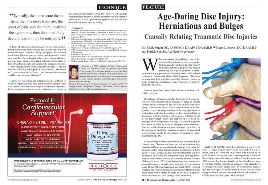
Age-Dating Disc Injury: Herniations and Bulges
FEATURE
Causally Relating Traumatic Disc Injuries
Mark Studin
William J. Owens
Patrick Sundby
DC, FASBE(C), DAAPM, DAAMLP,
DC, DAAMLP
Accident Investigator
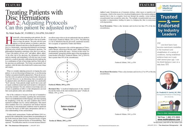
When considering disc pathology, one of the most asked questions is: How do you determine causality and age-date the lesion? Medical literature has purposefully dodged answering this type of question to apparently avoid any appearance of pandering to the medical-legal community. Fardon and Milette (2001) reported, “The term herniated disc does not infer knowledge of cause, relation to injury or activity, concordance with symptoms, or need for treatment” (p. E108).
Fourteen years later, with further evidence, Fardon et al. (2014) reported:
The category of trauma includes disruption of the disc associated with physical and/or imaging evidence of violent fracture and/or dislocation and does not include repetitive injury, contribution of less than violent trauma to the degenerative process, fragmentation of the ring apophysis in conjunction with disc herniation, or disc abnormalities in association with degenerative subluxations. Whether or not a “less than violent” injury has contributed to or been superimposed on a degenerative change is a clinical judgment that cannot be made on the basis of images alone; therefore, from the standpoint of description of images, such discs, in the absence of significant imaging evidence of associated violent injury, should be classified as degeneration rather than trauma, (p. 2531)
As described by Fardon, the definition and understanding of “violent injury” becomes an important arbiter in determining causality and lends an important understanding to age-date the herniation and/or bulge. In understanding the nature of Fardon’s tag of “violent,” science gives us answers rather than intuitive perceptions or rhetoric, and the quantification of energy transferences to victims in accidents gives us those answers. The rate of change in speed (AV) of any free-moving body contributes to quantifying energy transfer and can be directly correlated to injury. Brault, Wheeler, Siegmund, and Brault (1998) reported with rear-end collision testing, it was determined that whiplash could occur with a change in speed as low as 2.49 mph AV where there was no visual damage to the automobile.
Krafft et al. (2002) reported symptoms at a AV of 12.5 km (7.77 mph) and an injury mean threshold of 4.2 g for males, and a AV of 9.6 km (5.97 mph) with a mean of 3.6 g for females. Using this data, a corresponding window of time can be calculated between .084 seconds for males and .080 seconds for females (verifying that females are more at risk than males), resulting in a mean of .082 seconds. As evidenced previously, acceleration (AV) is important because it is part of the physics to determine g-forces, which explain injury thresholds and give a numerical value to Fardon et al. ’s (2014) use of the descriptor “violent injury.”
The risk for injury is present in a vehicle no matter the initial speeds or damage to the protective equipment because transference of forces is the prime factor in accidents. Additionally, low-speed collisions have a history of little to no damage. Therefore, little or no energy is absorbed by the safety equipment and design of the vehicles, yet the occupant is subject to these forces even with safety restraints. Because of these factors, only the following few pieces of information are needed to quantify the energy transfer an occupant is subjected to:
1. Speed just before the collision of each vehicle involved.
2. Weight of each vehicle and its occupants.
3. Time (t) of .082 seconds.
Two examples:
Example A: A 6,100 lb SUV traveling at 7 mph rear-ends a 4,200 lb car stopped at a red light, and the SUV stops as a result of the collision. The car (and its occupants) will experience a resultant AV of 10.67 mph (not to be confused with the speed of the bullet vehicle that struck the target vehicle).
Example B: A 30,000 lb truck and trailer backing up at 2 mph backs into the rear of a 4,800 lb occupied parked van, and the truck stops as a result of the collision. The van (and its occupants) will experience a AV of 12.5 mph.
AV AV Calculated G-Force (Miles per Hour) (Feet per Second) 10.67 14.95 5.66 12.5 18.38 6.96
Regarding Krafft et al.’s (2002) Tables 1 through 4 (Pg. 3): In both of the previous examples, the acceleration threshold for injury of males and females was exceeded. Both collisions would be traditionally classified as low speed with potentially no deformity of the vehicles. Just to underscore the injury potential at low speeds, the second example occurred at 2 mph where the physics of the crash offered demonstrable evidence of threshold forces sufficient to cause bodily injury.
Because Fardon et al. (2014) uses the word “violent” with no qualifying parameters, the previous examples offer insight through science about how transferred forces impact the human body with a predictable threshold for injury. Since the word “violent” is a subjective descriptor, one must utilize science and not consider generalities, as illustrated by the low-speed examples earlier.
Del Grande, Maus, and Carrino (2012) reported that, although there were varying reports of asymptomatic herniations in the literature, only a posttraumatic finding of radicular, or nerve root, pain can be definitive for determining causality.
Del Grande, Maus, and Canino (2012) wrote:
Only a close concordance, a key in lock fit, of an imaging finding and an individual patient’s pain syndrome can suggest causation, which further implies that the imager must know the nature of a radicular pain syndrome if he/she is to suggest a causal lesion. Close communication between clinician and imager via the medical record, an intake document at the imaging site detailing the pain syndrome, or direct patient interview by the imager is necessary, (p. 640)
Therefore, it is critical to ensure that patients have a complete history taken and an examination performed by a credentialed healthcare provider trained in trauma care. Many practitioners are licensed to treat the trauma case, but many are ill-equipped in training and experience to ensure an accurate diagnosis and determine proper relationship to causality.
Beyond radiating symptomatology, although as Del Grande, Maus, and Canino (2012) have reported as an accepted parameter for determining herniation causality, it is important to
‘ *The definition and understanding of “violent injury” becomes an important arbiter in determining causality and lends an important understanding to age-date the herniation and/or bulge. JJ
realize that radiating clinical symptoms arising out of injury to an intervertebral disc are dependent on the anatomical positioning of the injured and inflamed disc material. When the disc herniation is only of a lateralized nature, then the segmental nerve root is compressed or inflamed, producing radiation of axial symptoms to the corresponding upper or lower extremity. By discussing radiation as a primary indicator of acute traumatic injury to the intervertebral disc, central disc herniations are omitted, which do not typically produce extremity symptomatology. When it comes to acute injury in the absence or radiating symptoms, local symptomatology should also be considered in approaching a mechanism and timing of the injury. Furthermore, one must also look at the morphology or architecture of the individual vertebrate as demonstrative evidence to age-date disc pathology inclusive of both herniations and traumatically induced directional, nondiffuse bulges, as described by Fardon et al. (2014).
Wolff’s law is described by Isaacson and Bloebaum (2010) as, “Physical forces exerted on a bone, alter bone architecture
and is a well-established principle...” (p. 1271). This has been understood and accepted as a general principle since the late 1800s and has been verified through the past century’s research inclusive of contemporary research. Simply put, if a bone has abnormal stresses, it will change morphology or shape within expected parameters. Since these changes are “expected,” the question becomes: How does Wolff’s law apply to traumatic external forces and acute disc injury, and how does this relate to causality?
^Bone remodeling is a tightly coupled functional system and is strongly influenced by age, activity level, and mechanical loading. 55
In order to fully understand the process, it is critical to understand the biochemical reaction (functional adaptation) that occurs with abnormal stresses on bone, which centers on bioelectric changes that occur at the cellular level. According to Issacson and Bloebaum (2010), when tissue is damaged, the injury potential creates steady local electric fields that result from ion flux (positive and negative charges moving through local cellular membranes), which is an integral part in the regeneration/remodeling of boney tissue. Bone remodeling is a tightly coupled functional system and is strongly influenced by age, activity level, and mechanical loading. This functional adaptation of bone demonstrates the unique ability of bone to alter its trabecular (structural bone tissue) orientation because of loading conditions. According to Frost (1994), bone remodeling is a direct response to mechanical influences and strains on the osseous system. This can occur as a normal process to strengthen bone or as a response to altered anatomy, biomechanics, or direct traumatic injury. Since this is a predicable scenario, we can identify specific factors that will help us determine whether the response was present over time or is at the beginning phase of remodeling. That is the fundamental basis for putting a causally related date to the injury.
Isaacson and Bloebaum (2010) note that in regard to the remodeling of bone, the successful growth of additional supporting bone results from a combination of competent mechanical strain stimuli and endogenous electrical currents (bioelectrical changes). Simply, mechanical stresses and the flow of the bioelectric compounds work in conjunction with one another to strengthen or produce additional bone to functionally “buttress” the joint segment. The previously mentioned endogenous electrical current/bioelectrical changes are more commonly known as the piezoelectricity or the body’s electrical reaction to pressure or mechanical stress. It is this electrically and mechanically based system that subsequently controls osteogenic (osteo = bone, genic = to create) activity. The amplitude or amount of electrical potential is dependent on the magnitude of the mechanical bone loading, while polarity (application of the bioelectric charge) was determined by the direction of the
deformed bone. Isaacson and Bloebaum (2010) reported, “The specific loading pattern of bone has been documented as an important piezoelectric parameter since potential differences in bone have been known to be caused by charge displacement during the deformation period” (p. 1271). This means that the application of Wolff’s law to a boney segment is dependent on the amount of mechanical stresses as well as the direction of those forces, and therefore is based on basic engineering principles in the body. The extent and direction of the bone’s response to these forces is predictable and expected.
Additionally, Isaacson and Bloebaum (2010) noted that increased pressure surrounding the bone inhibits specific hormones, which prevents the uptake of calcium in the blood, and that, in turn, results in the additional uptake of calcium within bone itself, causing additional bone to be produced. Now that we understand what is happening from a physiological perspective when the bone responds to normal or abnormal mechanical stresses, the aging processes, or an acute traumatic injury, the question becomes: Can we objectively predict this process in the human spine?
He and Xinghua (2006) studied the predictability of bone remodeling, which included both the external shape and internal bone density distribution. They extended the simulation of the external shape of bones to determine and predict pathological changes in bone, specifically the osteophyte on the edge of
a bone structure. They reported, “The significance of this work were: ( 1) it confirmed that osteophyte formation was an adaptive process in response to the change of mechanical environment, which can be simulated numerically by combining quantitative bone remodeling theory with finite element method. And (2) it can help to better understand the relationship between bone morphological abnormity and the mechanical environment” (He and Xinghua, 2006, p. 96).
He and Xinghua (2006) also reported that with load-bearing bones, such as the femur and vertebrae, mechanical factors are crucial to the morphology and changes in boney structures, which relate closely to changes in mechanical environments. In addition, changes in bone-structure morphology are slowly progressing processes unless other factors, such as trauma or inflammation, are included, at which time the processes will be accelerated to change the bone structural morphology. This means that there is a “genetic timing” to the remodeling process that can be altered (increased) by the presence of specific conditions, such as an acute injury or inflammation.
According to He and Xinghua (2006), when only the local mechanical environment changes or a directional change in force coefficients is present, then only part of the vertebrate will remodel, leaving the rest of the vertebrate unchanged. Simply put, if there is a one-sided lesion creating pressure unilaterally, then only that side of the disc will create an osteophyte. This is very similar to the formation of a callus on your hand or foot. “In this paper, the main pathology of osteophyte formation was associated with the structural deterioration of intervertebral disc” (He and Xinghua, 2006, p. 97).
These researchers further discuss that the remodeling process will continue until the biomechanical failure is resolved and the body has reached a biomechanical equilibrium by placing an osteophyte on the edge of the vertebrate, one whose size and strength is based on the influencing mechanical imbalance. They concluded that only the bone in the area of mechanical imbalance would be compromised.
Although different individuals have different formation rates and the osteophytes may vary in size, everyone is subject to morphological changes depending on mechanical imbalances in the spine. He and Xinghua (2006) concluded that “... it will actually take about more than half a year to observe the bone morphological changes...” (p. 101). This indicates that it takes approximately six months for an osteophyte to be demonstrable after mechanical failure or imbalance. This
again gives a time frame to better understand if pathology of the intervertebral disc has been present for a long period (preexisting) or has been produced as the direct result of the specific traumatic event by lack of the existence of an osteophyte, meaning the disc pathology is less than six months in duration.
In conclusion, we would like to remind the reader that, by definition, a disc is a ligament, connecting a bone to a bone, and it has the structural responsibility to the vertebrate above and below it to keep the spinal system in equilibrium. Damage to the disc through a tear (herniation or annular fissure) or a directional, nondiffuse bulge will create abnormal load bearing or biomechanical failure on the side of the disc lesion. We have previously defined the term “violent trauma” as not being dependent on the amount of damage done to those structures either around or containing the victim, and have determined there were ample force coefficients to produce injury to the spine. So based on the current literature, we can now accurately predict in a demonstrable manner the timing of causality of the disc lesion. This is based on the symptomatology of the patient and/or the morphology of the vertebral structure, and is a subject that can no longer be based on rhetoric.
References:
1. Fardon, D. F, & Milette, P. C. (2001). Nomenclature and classification of lumbar disc pathology: Recommendations of the combined task forces of the North American Spine Society, American Society of Spine Radiology,
and American Society of Neuroradiology. Spine, 26(5), E93-E113.
2. Fardon, D. F„ Williams, A. L., Dohring, E. J., Murtagh, F. R., Rothman, S. L. G., & Sze, G. K. (2014). Lumbar Disc Nomenclature: Version 2.0: Recommendations of the combined task forces of the North American Spine Society, American Society of Spine Radiology, and American Society of Neuroradiology. Spine, 14(11), 2525-2545.
3. Brault J. R., Wheeler J. B., Siegmund, G. R, & Brault, E. J. (1998). Clinical response of human subjects to rear-end automobile collisions. Archives of Physical Medicine and Rehabilitation, 79(1), 72-80.
4. Krafft, M„ Kullgren, A., Malm, S., and Y denius, A. (2002). Influence of crash severity on various whiplash injury symptoms: A study based on real life rear-end crashes with recorded crash pulses. In Proc. 19th Int. Techn. Conf. on ESV, Paper No. 05-0363, 1-7.
5. Del Grande F„ Maus T. P, & Carrino J. A. (2012). Imaging the intervertebral disc: Age-related changes, herniations and radicular pain. Radiological Clinic of North America, 50(4), 629-649.
6. Issacson, B. M., & Bloebamn, R. D. (2010). Bone electricity: What have we learned in the past 160 years? Journal of Biomedical Research, 95 A(4), 1270-1279.
7. Frost, H. M. (1994). Wolff's Law and bone's structural adaptations to mechanical usage: an overview for clinicians. The Angle Orthodontist, 64(3), 175-188.
8. He, G., & Xinghua, Z. (2006). The numerical simulation of osteophyte formation on the edge of the vertebral body using quantitative bone remodeling theory. Joint Bone Spine, 73(1), 95-101.
Dr. Mark Studin is an adjunct associate professor o f chiropractic at the University of Bridgeport College of Chiropractic and an adjunct professor of clinical sciences at Texas Chiropractic College. He is also a clinical presenter for the State of New York at Buffalo; School of Medicine and Biomedical Sciences for
postdoctoral education, teaching MRI spine interpretation and triaging trauma cases. Dr. Studin is the president of the Academy of Chiropractic, teaching doctors of chiropractic how to interface with the legal community (www.DoctorsPIProgram.com); teaches MRI interpretation and triaging trauma cases to doctors of all disciplines nationally; and studies trends in health care on a national scale (www.TeachDoctors.com). Dr. Studin can he reached at [email protected] or at 631-786-4253.
Dr. Bill Owens is presently in private practice in Buffalo and Rochester, New York and generates the majority of his new patient referrals directly from the primary care medical community. He is an associate adjunct professor at the State University of New York
at Buffalo School of Medicine and Biomedical Sciences as well as the University of Bridgeport, College of Chiropractic and an adjunct professor of clinical sciences at Texas Chiropractic College. He also works directly with doctors of chiropractic to help them build relationships with medical providers in their community. He can he reached at [email protected], via www.mdreferralprogram.com, or by calling 716-228-3847.
automotive enforcement Patrick Sundby industry, collision has including investigation. decades several of He experience years has in also been in law the a driver training andfirearms instructor in law enforcement, as well as a police officer for nine years before
specializing in accident investigations. He has had the privilege participating in both learning and teaching at Prince William County Criminal Justice Training Academy in Virginia and studied at the Federal Law Enforcement Training Center in Georgia. His specialty is low-speed and catastrophic crashes and he has testified more than 500 times at various levels. He can be reached at 571-265-8076 or patrick. sundby@gmail. com.