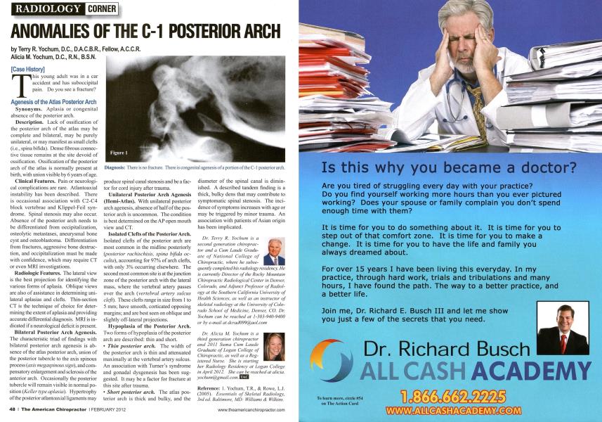
[Case History] T his young adult was in a car accident and has suboccipital pain. Do you see a fracture? Agenesis of the Atlas Posterior Arch Synonyms. Aplasia or congenital absence of the posterior arch. Description. Lack of ossification of the posterior arch of the atlas may be complete and bilateral, may be purely unilateral, or may manifest as small clefts (i.e.,spinabifida). Dense fibrous connective tissue remains at the site devoid of ossification. Ossification of the posterior arch of the atlas is normally present at birth, with union visible by 6 years of age. Clinical Features. Pain or neurological complications are rare. Atlantoaxial instability has been described. There is occasional association with C2-C4 block vertebrae and Klippel-Feil syndrome. Spinal stenosis may also occur. Absence of the posterior arch needs to be differentiated from occipitalization, osteolytic metastases, aneurysmal bone cyst and osteoblastoma. Differentiation from fractures, aggressive bone destruction, and occipitalization must be made with confidence, which may require CT or even MRI investigations. Radiologic Features. The lateral view is the best projection for identifying the various forms of aplasia. Oblique views are also of assistance in determining unilateral aplasias and clefts. Thin-section CT is the technique of choice for determining the extent of aplasia and providing accurate differential diagnosis. MRI is indicated if a neurological deficit is present. Bilateral Posterior Arch Agenesis. The characteristic triad of findings with bilateral posterior arch agenesis is absence of the atlas posterior arch, union of the posterior tubercle to the axis spinous process (axis megaspinoiis sign), and compensatory enlargement and sclerosis of the anterior arch. Occasionally the posterior tubercle will remain visible in normal position (Keller type aplasia). Hypertrophy of the posterior atlantoaxial ligaments may produce spinal canal stenosis and be a factor for cord injury after trauma. Unilateral Posterior Arch Agenesis (Hemi-Atlas). With unilateral posterior arch agenesis, absence of half of the posterior arch is uncommon. The condition is best determined on the AP open mouth view and CT. Isolated Clefts of the Posterior Arch. Isolated clefts of the posterior arch are most common in the midline posteriorly (posterior rachischisis, spina bifida oc-culta), accounting for 97% of arch clefts, with only 3% occurring elsewhere. The second most common site is at thejunction zone of the posterior arch with the lateral mass, where the vertebral artery passes over the arch {vertebral artery sulcus cleft). These clefts range in size from 1 to 5 mm; have smooth, corticated opposing margins; and are best seen on oblique and slightly off-lateral projections. Hypoplasia of the Posterior Arch. Two forms of hypoplasia of the posterior arch are described: thin and short. Thin posterior arch. The width of the posterior arch is thin and attenuated maximally at the vertebral artery sulcus. An association with Turner's syndrome and gonadal dysgenesis has been sug gested. It may be a factor for fracture at this site after trauma. Short posterior arch. The atlas pos terior arch is thick and bulky, and the diameter of the spinal canal is diminished. A described tandem finding is a thick, bulky dens that may contribute to symptomatic spinal stenosis. The incidence of symptoms increases with age or may be triggered by minor trauma. An association with patients of Asian origin has been implicated. Dr. Terry R. Yochum is a second generation chiropractor and a Cum Laude Graduate of National College of Chiropractic, where he subse- quently completed his radiology residency. He is currently Director of the Rocky Mountain Chiropractic Radiological Center in Denver, Colorado, and Adjunct Professor of Radiology at the Southern California University of Health Sciences, as well as an instructor of skeletal radiology at the University of Colorado School of Medicine, Denver, CO. Dr. Yochum can he reached at 1-303-940-9400 or bv e-mail at [email protected] Dr. Alieia M. Yochuin is a Ihird generation chiropractor and 2011 Snma Cum Lande Graduate of Logan College of Chiropractic, as well as a Registered Nurse. She is starting her Radiology Residency at Logan College in April 2012. She can he reached at alicia. [email protected]. EZS3 Reference: 1. Yochum, T.R., & Rowe, L.J. (2005). Essentials of Skeletal Radiology, irded. Baltimore, MD: Williams & Wilkins. Diagnosis: There is no fracture. There is congenital agenesis of a portion of the C-1 posterior arch.