Why Support the Three Arches of the Foot?
BIOMECHANICS
Kevin Wong
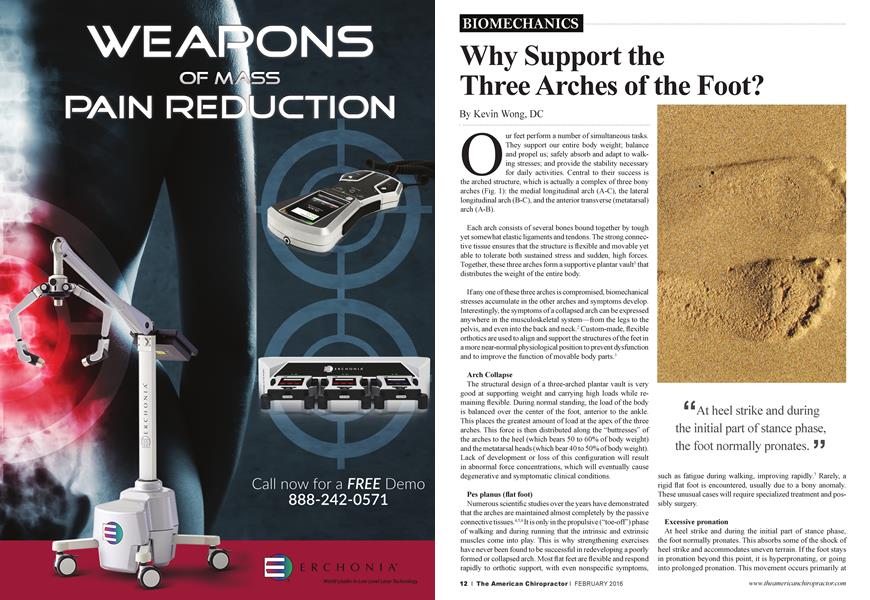
Our feet perform a number of simultaneous tasks. They support our entire body weight; balance and propel us; safely absorb and adapt to walking stresses; and provide the stability necessary for daily activities. Central to their success is the arched structure, which is actually a complex of three bony arches (Fig. 1): the medial longitudinal arch (A-C), the lateral longitudinal arch (B-C), and the anterior transverse (metatarsal) arch (A-B).
Each arch consists of several bones bound together by tough yet somewhat elastic ligaments and tendons. The strong connective tissue ensures that the structure is flexible and movable yet able to tolerate both sustained stress and sudden, high forces. Together, these three arches form a supportive plantai' vault1 that distributes the weight of the entire body.
If any one of these three arches is compromised, biomechanical stresses accumulate in the other arches and symptoms develop. Interestingly, the symptoms of a collapsed arch can be expressed anywhere in the musculoskeletal system—from the legs to the pelvis, and even into the back and neck.2 Custom-made, flexible orthotics aie used to align and support the structures of the feet in a more near-nonnal physiological position to prevent dysfunction and to improve the function of movable body parts. '
Arch Collapse
The structural design of a three-arched plantai' vault is very good at supporting weight and carrying high loads while remaining flexible. During normal standing, the load of the body is balanced over the center of the foot, anterior to the ankle. This places the greatest amount of load at the apex of the three arches. This force is then distributed along the “buttresses” of the arches to the heel (which bears 50 to 60% of body weight) and the metatarsal heads (which bear 40 to 50% of body weight). Lack of development or loss of this configuration will result in abnormal force concentrations, which will eventually cause degenerative and symptomatic clinical conditions.
Pes planus (flat foot)
Numerous scientific studies over the years have demonstrated that the arches aie maintained almost completely by the passive connective tissues.4’56 It is only in the propulsive (“toe-off’) phase of walking and during running that the intrinsic and extrinsic muscles come into play. This is why strengthening exercises have never been found to be successful in redeveloping a poorly formed or collapsed arch. Most flat feet aie flexible and respond rapidly to orthotic support, with even nonspecific symptoms,
‘ ‘At heel strike and during the initial part of stance phase, the foot normally prônâtes. J J
such as fatigue during walking, improving rapidly.7 Rarely, a rigid flat foot is encountered, usually due to a bony anomaly. These unusual cases will require specialized treatment and possibly surgery.
Excessive pronation
At heel strike and during the initial part of stance phase, the foot normally pronates. This absorbs some of the shock of heel strike and accommodates uneven terrain. If the foot stays in pronation beyond this point, it is hyperpronating, or going into prolonged pronation. This movement occurs primarily at
the subtalar and talonavicular joints, with excessive loading affecting all of the arches, although it affects the medial arch most acutely. Excessive pronation causes an obvious flattening of the medial longitudinal arch, with a medial and inferior movement of the navicular bone.
Navicular drop
A quick test that measures the change in position of the navicular prominence can be performed to quantify the presence of arch collapse during weight bearing.6 The navicular drop test is especially useful with runners,8 since it shows the change in arch height from non-weight bearing to weight bearing, as well as any asymmetry between left and right arches. The test helps to verify poor spinal support from the arches and demonstrates the need for orthotics to the patient. Foot Levelers, Inc. has developed the “Postural Stability Indicator” card to record the results of this test. No fancy equipment is necessary, and the small amount of time spent in testing for arch collapse is greatly rewarded.
What Can Ortho tics Do?
Static support. The foot and ankle provide support and balance for the body while absorbing shock.9 During standing posture, the alignment of the arches in each foot has a significant impact on the position of the legs and pelvis. When the arches ai e low and/or pronating excessively, the lower extremities tend to rotate medially. A recent study using radiographic measurements found that custom-made flexible orthotics can significantly improve the alignment of the arches when standing.10
Dynamic support
During gait, the foot undergoes substantial changes. The arches and connective tissues must sustain the stress of heel strike, then adapt to the ground during stance phase, and finally become a rigid lever to provide an efficient push-off. This must all occur in a coordinated manner, with no “glitches” or hang-ups. The foot must permit a smooth transfer of the body’s center of mass over the leg in order to conserve energy and keep the work expenditure to a minimum.11 And the heavier a patient is, the greater the stresses on the feet and ankles. This requires an orthotic to be flexible yet supportive, and orthotic designs must consider weight and intensity of forces, in addition to encouraging proper movement and function of the foot while supporting all three arches.
Postural benefits
Orthotics designed to provide support for the arches of the feet can have many additional benefits. Since the entire body structure is balanced on one foot at a time when walking and running, improving foot alignment can improve knee, hip, pelvis, and even spinal postural alignment. A low femur head seen on properly positioned postural films indicates a difference in leg length. While there ai e several causes (from injury to growth asymmetry to arch collapse), most patients will benefit from the additional support provided by a pair of orthotics.12 An added heel lift may also be necessary in some cases. Joint degeneration (of the hip, knee, or spinal joints) with wealing of the cartilage requires the additional support and shock absorption provided by orthotics. A pelvic or spinal tilt or recurrent subluxations will often respond rapidly to orthotic support of the arches in the feet.13
Conclusion
It’s not unusual to have musculoskeletal complaints in the legs, hips, and spine from malfunctioning arches. A brief screening exam can help identify the commonly seen clues, such as lowered arches, heel eversion, Achilles tendon bowing, and leg length differences. Well-designed, custom-fitted, flexible orthotics can provide much of the support that is lacking and can improve locomotor efficiency by guiding the calcaneus and arches through the gait cycle.
References
1. Kapandji IA. Physiology of the joints: Lower limb. 2nd ed. New York: Churchill Livingstone, 1981:154-182.
2. Posner, Anthony et al. Influence of foot orthotics upon duration of effects of spinal manipulation in chronic back pain patients: A randomized clinical trial. J Manip Physiol Ther 2014;37:124-140.
3. Ferrari Robert. Effect of customized foot orthotics in addition to usual care for the management of chronic low back pain following work-related low back injury. J Manip Physiol Ther 2013;6:359-363.
4. Basmajian JV, Bentzon JW. An electromyographic study of certain muscles of the leg and foot in the standing position. Surg Gynec & Obstet. 1954;98:662-666.
5. Basmajian JV, Stecko G. The role of muscles in arch support of the foot: An electromyographic study. J Bone Joint Surg. 1963;45-A: 1184-1190.
6. Huang C-K et al. Biomechanical evaluation of longitudinal arch stability. Foot & Ankle. 1993;14:353-357.
7. Otman S et al. Energy cost of walking with flat feet. Prosth Orthot Interntl. 1988;12:73-76.
8. Gould N. Evaluation of hyperpronation andpes planus in adults. Clin Orthop. 1983;181:37-45.
9. Brody D. Techniques in the evaluation and treatment of the injured runner Orthop Clin North Am. 1982; 13:541-558.
10. Souza TA. Differential diagnosis for the chiropractor: Protocols and algorithms. Gaithersburg, MD: Aspen Publishers, 1997:323.
11. Kuhn DR etal. Radiographic evaluation of weight-bearing orthotics and their effect on flexible pes planus. J Manip Physiol Ther. 1999;22:221-226.
12. Kirby KA, Biomechanics of the normal and abnormal foot. J Am Podiatr Med Assoc. 2000;90:30-34.
13. Bay lis WJ, Rzonca EC. Functional and structural limb length discrepancies: Evaluation and treatment. Clin Podiatr Med Sur. 1988;5:509-520.
14. Rothbart BA, Estabrook L. Excessive pronation: A major biomechanical determinant in the development of chondromalacia and pelvic lists. J Manip Physiol Ther. 1988;11:373-379.
Dr. Kevin Wong is a 1996 summa cum laude graduate of Palmer College of Chiropractic West. A past instructor of chiropractic technique for the college, he is currently part of the adjunct faculty. Dr Wong is also a past lecturer and instructor for the International Chiropractic Association of California. Currently he is in full-time practice in Orinda, California, and has been a speaker for Foot Levelers for many years.