Pronation, Posture and Piriformis Syndrome
BIOMECHANICS
Putting the foot down on sciatica
Jennifer Illes
BSc, DC, MS
There is a direct and evidence-based relationship between movements of the lower extremity and associated stability of the pelvis and spine. A link has even been uncovered between low back pain and the subsequent development of cervical symptoms, suggesting a chain of reactions throughout the musculoskeletal system.1 For a healthy patient, we must help establish patterns of joint and muscular interactions that function automatically, from the ground upward (i.e., following the kinetic chain). Proper joint function can be affected by gait, excessive pronation, leg length inequality (LLI), and uncoordinated muscle patterns. These factors may be overlooked obstacles in the proper treatment of joint restrictions and pain syndromes, including sciatica.2
Sciatica is a debilitating condition in which the patient experiences pain and/or paresthesias in the distribution of the sciatic nerve or an associated lumbosacral nerve root. Often, a common mistake is referring to any low back pain or radicular leg pain as sciatica. The sciatic nerve is made up of nerve roots/rootlets from L4 to S2.
Sciatica pain often is worsened with flexion of the lumbar spine, twisting, or coughing. It provides direct motor function to the hamstrings, lower extremity adductors, and indirect motor function to the calf muscles, anterior lower leg muscles, and some intrinsic foot muscles. Although there are no direct sensory functions of the sciatic nerve, it indirectly innervates (via its terminal branches) the skin of the lateral leg, heel, and both the dorsal and plantar surfaces of the foot. Some newer research has identified highfrequency vibration losses for those with chronic forms of sciatica.3
What to Look For
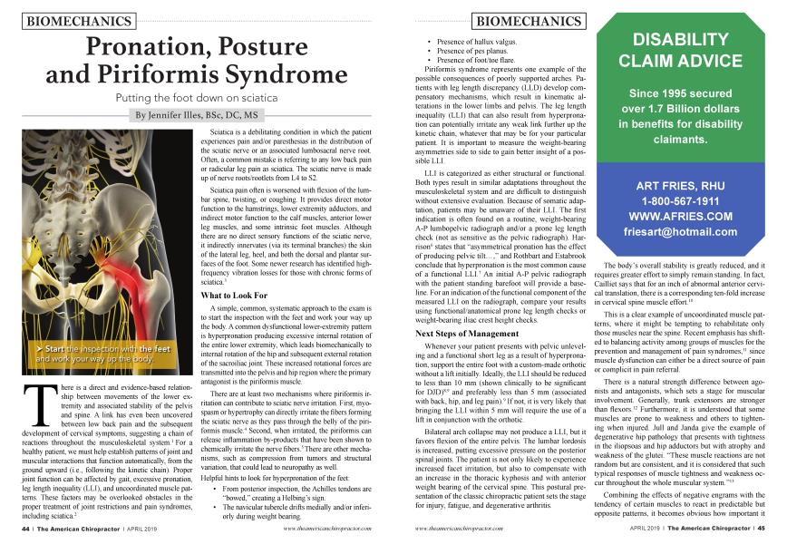
A simple, common, systematic approach to the exam is to start the inspection with the feet and work your way up the body. A common dysfunctional lower-extremity pattern is hyperpronation producing excessive internal rotation of the entire lower extremity, which leads biomechanically to internal rotation of the hip and subsequent external rotation of the sacroiliac joint. These increased rotational forces are transmitted into the pelvis and hip region where the primary antagonist is the piriformis muscle.
There are at least two mechanisms where piriformis irritation can contribute to sciatic nerve irritation. First, myospasm or hypertrophy can directly irritate the fibers forming the sciatic nerve as they pass through the belly of the piriformis muscle.4 Second, when irritated, the piriformis can release inflammation by-products that have been shown to chemically irritate the nerve fibers.5 There are other mechanisms, such as compression from tumors and structural variation, that could lead to neuropathy as well.
Helpful hints to look for hyperpronation of the feet:
• From posterior inspection, the Achilles tendons are “bowed,” creating a Helbing’s sign.
• The navicular tubercle drifts medially and/or inferiorly during weight bearing.
• Presence of hallux valgus.
• Presence of pes planus.
• Presence of foot/toe flare.
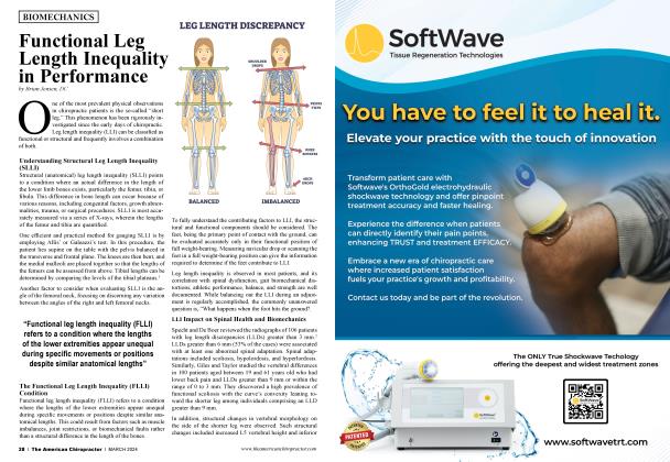
Piriformis syndrome represents one example of the possible consequences of poorly supported arches. Patients with leg length discrepancy (LLD) develop compensatory mechanisms, which result in kinematic alterations in the lower limbs and pelvis. The leg length inequality (LLI) that can also result from hyperpronation can potentially irritate any weak link further up the kinetic chain, whatever that may be for your particular patient. It is important to measure the weight-bearing asymmetries side to side to gain better insight of a possible LLI.
LLI is categorized as either structural or functional. Both types result in similar adaptations throughout the musculoskeletal system and are difficult to distinguish without extensive evaluation. Because of somatic adaptation, patients may be unaware of their LLI. The first indication is often found on a routine, weight-bearing A-P lumbopelvic radiograph and/or a prone leg length check (not as sensitive as the pelvic radiograph). Harrison6 states that “asymmetrical pronation has the effect of producing pelvic tilt...,” and Rothbart and Estabrook conclude that hyperpronation is the most common cause of a functional LLI.7 An initial A-P pelvic radiograph with the patient standing barefoot wifi provide a baseline. For an indication of the functional component of the measured LLI on the radiograph, compare your results using functional/anatomical prone leg length checks or weight-bearing iliac crest height checks.
Next Steps of Management
Whenever your patient presents with pelvic unleveling and a functional short leg as a result of hyperpronation, support the entire foot with a custom-made orthotic without a lift initially. Ideally, the LLI should be reduced to less than 10 mm (shown clinically to be significant for DJD)8-9 and preferably less than 5 mm (associated with back, hip, and leg pain).9 If not, it is very likely that bringing the LLI within 5 mm wifi require the use of a lift in conjunction with the orthotic.
Bilateral arch collapse may not produce a LLI, but it favors flexion of the entire pelvis. The lumbar lordosis is increased, putting excessive pressure on the posterior spinal joints. The patient is not only likely to experience increased facet irritation, but also to compensate with an increase in the thoracic kyphosis and with anterior weight bearing of the cervical spine. This postural presentation of the classic chiropractic patient sets the stage for injury, fatigue, and degenerative arthritis.
The body’s overall stability is greatly reduced, and it requires greater effort to simply remain standing. In fact, Cailliet says that for an inch of abnormal anterior cervical translation, there is a corresponding ten-fold increase in cervical spine muscle effort.10
This is a clear example of uncoordinated muscle patterns, where it might be tempting to rehabilitate only those muscles near the spine. Recent emphasis has shifted to balancing activity among groups of muscles for the prevention and management of pain syndromes,11 since muscle dysfunction can either be a direct source of pain or complicit in pain referral.
There is a natural strength difference between agonists and antagonists, which sets a stage for muscular involvement. Generally, trunk extensors are stronger than flexors.12 Furthermore, it is understood that some muscles are prone to weakness and others to tightening when injured. Jull and Janda give the example of degenerative hip pathology that presents with tightness in the iliopsoas and hip adductors but with atrophy and weakness of the glutei. “These muscle reactions are not random but are consistent, and it is considered that such typical responses of muscle tightness and weakness occur throughout the whole muscular system.”13
Combining the effects of negative engrams with the tendency of certain muscles to react in predictable but opposite patterns, it becomes obvious how important it is to evaluate our patients’ global posture. The most effective adjustments may be diminished through uncooperative muscle activity.
Improperly functioning muscles also affect normal transmission of proprioceptive feedback, another mechanism in the kinetic chain relationship. “The importance of adequate sensory input, proprioceptive control and proper function of sensorimotor integration has probably been underestimated in the pathogenesis of low back pain... For this reason, proprioceptive facilitation techniques should be included in the therapeutic programs for those suffering from low back pain syndromes and postural defects.”13
Chiropractic adjustments of the spine improve proprioceptive input by normalizing joint alignment and muscle tonus. The manipulation helps with dysafferentation. This means that it increases mechanoreceptor activity and decreases nociceptor activity. Furthermore, because the feet contain approximately one-quarter of all the body’s joints and, therefore, a concentration of proprioceptive fibers, it becomes logical to conclude that support of the postural foundation using custom-made orthotics will enhance balance and muscle coordination. In fact, this was the conclusion reached during research involving custom orthotics, published in the Journal of Manipulative and Physiological Therapeutics.14
The reason a diagnosis of true sciatic nerve irritation can be difficult to reach is the comprehensive nature of the structures involved in the pathogenesis of this condition. Chiropractors are best equipped to address the several components of joint dysfunctions, muscular imbalance, and sensorimotor feedback errors. However, unless a global approach is taken for this and most other conditions, results will be short-lived. Successful treatments will combine specific adjustments to counteract the patterns of joint dysfunction, combined with rehabilitation of specific muscle groups and support for deficient structures, including the feet and lower extremities.
References
1. Horal J. The clinical appearance of low back disorders in the city of Gothenburg, Sweden. Acta Orthop Scand suppl. 118:15, 1969.
2. Yekutiel MP. The role of vertebral movement in gait: implications for manual therapy. J Man Manip Ther. 1994; 2:22-27.
3. Bijman M, Strzalkowski ND, Bent LR, Brown SH. Deficits in foot skin sensation are related to alterations in balance control in chronic low back patients experiencing clinical signs of lumbar nerve root impingement. Frost LR, Gait Posture. 2015 May;41(4):9238. doi: 10.1016/j.gaitpost.2015.03.345.
4. Cox JM. Low Back Pain: Mechanism, Diagnosis and Treatment, 5th ed. Baltimore: Williams & Wilkins, 1990.
5. Steiner C et al. Piriformis syndrome: pathogenesis, diagnosis, and treatment. J Am Osteopath Assoc. 1987; 87:318. Harrison, D. etal. CPB, Vol. 4. 1998.
6. Giles LGF, Taylor JR. Lumbar spine structural changes associated with leg length inequality. Spine. 1982;7:159-162.
7. Giles LGF, Taylor JR. Low back pain associated with leg length inequality. Spine. 1981; 6:510-521.
8. Friberg O. Clinical symptoms and biomechanics of lumbar spine and hip joint in leg length inequality. Spine. 1983, 6(6): 643-650.
9. Walsh M Connolly P, Jenkinson A, O’Brien T. Leg length discrepancy: an experimental study of compensatory changes in three dimensions using gait analysis. Gait Posture. 2000 Oct;12(2): 156-61.
10. Cailliet R. Neck and Arm Pain. Philadelphia: F.A. Davis, 1981.
11. Lewit K. Manipulative Therapy in Rehabilitation of the Motor System. Butterworth, London, 1985.
12. Langrana NA, Lee CK, Alexander H, Mayott CW. Quantitative assessment of back strength using isokenetic testing. Spine. 9:287, 1984.
13. Jull GA, Janda V. Muscles and Motor Control in Low Back Pain: Assessment and Management. In Physical Therapy of the Low Back. Churchill Livingstone, New York 1987.
14. Stude DE, Brink DK. Effects of nine holes of simulated golf and orthotic intervention on balance and proprioception in experienced golfers. J Manip Physiol Ther. 1997; 20:590-601.
Dr. Hies is a dynamic communicator who brings 15 years of combined clinical and academic experience. She is a graduate of New York Chiropractic College and her experience includes treating professional athletes with sports related injuries. She is an expert on extremity adjusting and is passionate about educating healthcare physicians. In her seminar, Dr. Hies shows how to complete a proper head and neck examination, how to diagnose common TMJ dysfunction, and effective rehabilitation exercises and techniques. Dr. Hies currently serves as an associate professor at Keiser University's College of Chiropractic Medicine in West Palm Beach, FL.