The topic of plastic deformation of the foot’s support structures is often discussed when evaluating feet for signs of excessive pronation as it applies to posture, gait, and functional movement. By definition, “Plastic deformation is the permanent distortion that occurs when material is subjected to tensile, compressive, bending, or torsion stresses that exceed its yield strength and cause it to elongate, compress, buckle, bend, or twist.”1
In a study of the anatomy of the plantar fascia2, it is interesting to note that the majority of the fibers are collagen and are oriented in a longitudinal direction, which is consistent with aponeurosis. However, there are also multidirectional fibers lying in vertical, transverse, and oblique directions, which are characteristic of fascia. The terms plantar fascia and plantar aponeurosis are used interchangeably.
The plantar fascia is richly innervated, especially where it joins with the myofascial of the abductor hallucis and abductor digiti minimi. Together with the flexor hallucis longus and flexor hallucis brevis muscles, abductor hallucis aids the flexion of the big toe. The muscle also helps maintain the medial longitudinal arch of the foot while walking. The abductor hallucis muscle is innervated by the medial plantar nerve (SI to S3), the larger of the two terminal branches of the tibial nerve. Pacini and Ruffini corpuscles have also been observed, suggesting that the plantar fascia has a role in proprioception, stability, and peripheral motor control of foot movements.
The fundamental role of the plantar fascia is to support the medial, lateral, and transverse arches of the foot, dissipate the shock forces generated during gait and provide propulsion from the stored strain energy in the tissue.
Researchers were surprised to find hyaluronic acid inside the plantar fascia, which is present when repair and regeneration of tissue occurs. This suggests the plantar fascia undergoes a form of pathological degeneration commonly referred to as plantar fasciitis, which is the inflammatory response to small tears in the collagen fibers. Elastic fibers make up a small amount of the plantar fascia content, but little is known about its extracellular matrix or innervation.
In the 1950s, chiropractors became aware of the importance of plantar fascia and its function after Dr. Monte Greenawalt observed how excessive pronation impacted the effectiveness of his treatment. He noticed that the pedal foundation often allowed the foot to pronate excessively and asymmetrically between the right and left foot. This deformation activated nociceptive mechanoreceptors, inhibiting postural tone, creating changes in normal gait, and in some cases, activating the pain reflex.
Adjustments to the arches of the feet and exercises to strengthen the intrinsic muscles of the feet to restore the arches failed to make any long-lasting changes, indicating that the loss of the arch structure was permanent.
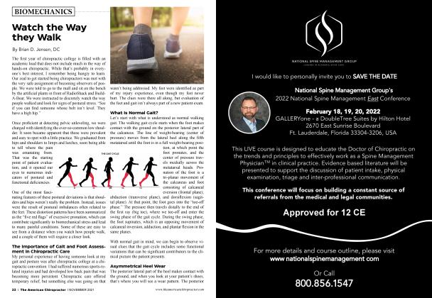
Repeated stretching of the plantar fascia from walking, running, jumping, and even standing on hard surfaces is enough to create the microtrauma to the collagen fibers that causes the arches to lose their integrity. The fact that the tissue does not return to its original length indicates that the force has exceeded its yield strength and caused it to elongate permanently. The fascia/aponeurosis does not have the ability to contract back to its original length, causing permanent plastic deformation.
The development of three-dimensional mapping and scanning technology has given us the tool to evaluate our chiropractic patients to determine the integrity of the plantar vault. Observational studies have revealed that the plantar fascia is deficient in its ability to keep the foot in an optimal structural and functional position in nearly 80% of our patient base.
Optimizing the function of the plantar fascia with a custom orthotic that supports all three arches in their optimal position effectively blocks excessive pronation and allows the plantar fascia to activate the proprioceptive functions present in the foot. This optimized state has been observed to change functional movement and posture and has resulted in countless positive patient testimonials. When the arches of the feet function within optimal ranges of motion, the mechanoreceptors activate efficient muscle responses in the lower extremity and enhance the effect of the chiropractic adjustment, which allows patients to move efficiently with stronger posture.
Dr. Brian Jensen graduated from Palmer College of Chiropractic in 1987. He is a Foot Levelers speaker. Dr. Jensen has his own practice in Roanoke, VA.
References:
1. ScienceDirect, Materials Enabled Designs, 2009
2. Journal of Anatomy, 2013 Dec; 223(6):665-676