Ankle and Foot Strains: A Comprehensive Approach
By Stephan Ediss, DC, PAK, FIACA
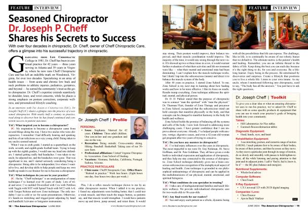
When it comes to athletic injuries, one of the biggest game-changers is an ankle or foot sprain/strain. Although sprains and strains of the ankle and foot can happen frequently while participating in sports, people can suffer that injury by rolling an ankle just walking down the street. In this article, we will review the mechanism of failure for the ankle joint, an assessment protocol for thorough evaluation, nutritional support, and therapeutic techniques to expedite recovery from an ankle injury.
The mortise joint is one of the most important joints of our entire body. It is the gateway for proprioceptive input during weight-bearing activities. When left untreated, an ankle injury can be a source for chronic and recurring issues for the body on the same side as the injured ankle for years to come. This occurs because of negative proprioceptive feedback from the injured joint. As an example, a chronic knee, sacroiliac joint, or rotator cuff problem may become an issue because of chronic negative proprioceptive feedback from an old injury to the mortise joint.
What is unique about the anatomy of the mortise joint is that it is the only joint in the human body with no muscular attachments. It is held together solely by a network of ligaments armed with Golgi tendon organs (GTOs). These GTOs are designed to protect the joint and the surrounding bony structures in the event of improper mechanics or mechanical overload. Once the mortise joint reaches a critical angle of position or ROM that compromises its structures, the GTOs will inhibit the supporting muscles and tendons, allowing the ankle to fail in an attempt to protect it from even greater damage.
For example, in the event of an inversion ankle sprain, once the ankle joint reaches a critical angle position, the GTOs will inhibit the peroneus brevis and longus, allowing the ankle to continue laterally. This reflexive response is designed to spare the tendons and muscles involved, although some injury to ligaments and tendons that support the lateral aspect of the ankle will still occur, with hopefully less overall severity. As the lateral structures stretch, the medial structures may compress. The result is circumferential swelling and bruising.
Following an event like that, it is recommended that proper precautionary screening is warranted, using standard methods of orthopedic testing and diagnostic imagery to rule out fractures and dislocations. Once it has been established that it is safe to proceed, this treatment procedure has proven to be safe, effective, and relatively quick, resulting in a more complete and faster recovery.
The first step is to “delete” the injury. With the patient seated with their legs hanging comfortably, the doctor applies gentle traction as equally around the mortise joint as possible, combining very slight inversion and eversion motion (approximately 8 oz, three to five seconds).
Immediately following, the doctor uses the web of their hand over the neck of the talus. With the fingers of the same hand, the doctor applies upward pressure to the bottom of the forefoot, creating a very light dorsiflexion rocking motion, which will reverse the activated withdrawal reflex from the original injury. This injury recall technique (IRT) will basically “delete” a bad file (the injury itself) from the nervous system’s memory bank. When performed properly, mechanical loading will no longer inhibit ipsilateral muscular function.
The next step is to deactivate the GTOs involved. This is easily achieved by applying enough digital pressure on the injured ligaments to stretch them and then tap a convenient DTR (e.g., patella tendon). Challenge the effectiveness of the correction by putting weight through the mortise joint and muscle-response testing (a strong muscle test should remain strong while putting weight through the mortise joint or rubbing the injured soft tissues).
Key nutritional support to the injured muscles and inflammation is the last key component for a speedy recovery. The muscles of the lower leg typically fail, not only because of the inhibitory response of the nervous system, as previously mentioned, but also due to adrenal fatigue, depletion of necessary minerals, and dehydration. Supplement choices should have all necessary nutritional components designed in a synergistic fashion to provide core base nutrients as well as their co-factors for maximum absorption and use. For accuracy, the injured, weakened muscles should demonstrate restored function when testing the patient (holding or in-salivating the product of choice).
Finally, the resolution of inflammation comes from a three-pronged approach. Resetting the mechanoreceptors decreases aberrant signals from the nervous system, thus addressing the inflammatory response. Secondly, because of the potential harmful side effects of various OTC pain medications, proteolytic plant enzymes can be a highly effective alternative. Last but certainly not least, the application of a topical has clinically proven to be effective in addressing pain, inflammation, and bruising. The one I use in my clinic contains proteolytic enzymes as well as homeopathic remedies known for injury support.
Please bear in mind that, although the above information and techniques are highly effective when treating sprains and strains of the foot and ankle, we still want to combine these techniques with other appropriate modalities and restorative techniques, such as manipulation, various physiotherapy modalities, compression, ice, etc.
About the Author
Stephan P. Ediss, DC, PAK, FIACA, has over 300 hours of certified training as a professional applied kinesiologist and has been in practice for 31 years. All his knowledge is compiled in an organized, easy-to-follow, step-by-step protocol, resulting in amazing clinical outcomes. Call him at 307-358-3147 or email at [email protected].