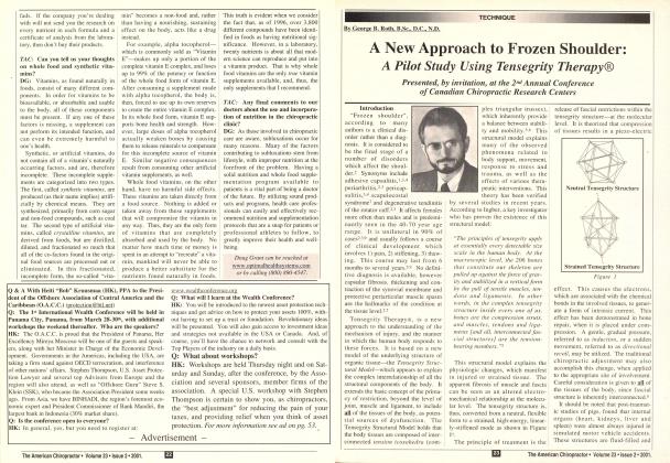
"Jhe word 'tenscgritv' is an invention: a contraction of 'tensional integrity.' Ten-segrity describes a structural-relationship principle in which structural shape is guaranteed by the finitely closed, comprehensively continuous, tensional behaviors of the system and not by the discontinuous and exclusively local compression member behaviors. Ten-segrity provides the ability to yield increasingly without ultimately breaking or coming asunder." - Buckminster Fuller . The spine has been the subject of endless study and debate. Back pain is a universal complaint and scientists and clinicians have probed, imaged, dissected, fused, discectomized, injected, bolted, inserted rods, pummeled, cracked, stretched, twisted and exer-cised-ad infinitum-in the elusive attempt to alleviate this age-old affliction. The advent of early imaging techniques, namely the X-ray, presented the spine as a disembodied stack of bones, separated by essentially invisible (to the X-ray) discs and other soft tissues. The apparent misalignment of vertebrae and/or the collapse of the intervening space, led to the conclusion that this "fragile" and frequently imbal-anced structure must be the source of the patient's symptoms.6 Indeed the spine is richly supplied with pain and kinesthetic receptors of every description, and stimulation of these pain receptors will reproduce the presenting complaint. The ultimate medical intervention to relieve this condition has been to ablate the offending pain nerve fibers.2 Nothing like treating the cause! A UNIFIED STRUCTURAL MODEL Theories regarding the function of the musculos-keletal system have borrowed piecemeal from mechanical engineering principles, which were originally developed to describe the characteristics of man-made materials and structures. Columns, posts and lintels, hinges, struts, suspension braces, guy wires, pulleys and levers have variously been applied to living organisms with limited success and even less consistency. Conventional engineering models apply to one or several of the characteristics associated with organic forms, but cannot be universally applied."^ The need for a unified model, applicable to living structures, has important clinical implications. The principles which govern the intrinsic nature of living tissue should, in theory, form the basis of restoration of function. The fact that there exist so many divergent therapeutic models is a testament to the veritable Babylon of structural concepts upon which they are based. The various therapeutic systems each have some value within certain applications, but appear to be limited in others. This fact would support the conclusion that the structural model upon which they are based must, in some way, be deficient. The concept of Tensegrity. or the Ten-segrity Structural Model (TSM), as a viable model to explain the complex properties of living organisms was put forward by Stephen M. Levin, M.D. This model, which is essentially an icosohedral crystalline extension of the carbon atom, appears to answer many of the seemingly impossible functional characteristics of organic forms. It is inherently stable and independent of gravitational forces and has properties to allow for controlled movement and the transmission of forces throughout the entire contiguous structure of the organism. This model explains the tremendous forces required to support vertical weight-bearing, as well as the horizontal forces necessary to allow for a giraffe to bend over for a drink of water and for a gymnast to project her body over seemingly impossible distances and bend into extreme contortions. Many of these postures and movements would cause a column supported by guy wires to literally fly apart.3 The Tensegrity Structural Model has now been verified as the underlying structure of all organic tissue.6 It explains many of the observed phenomena related to body support, movement, response to stress and trauma; as well as the effects of various therapeutic interventions. This theory has been verified by several studies in recent years. According to Ingber, a key investigator who has proven the existence of this structural model: "The principles of tensegrity apply at essentially every detectable size scale in the human body. At the macroscopic level, the 206 bones that constitute our skeleton are pulled up against the force of gravity and stabilized in a vertical form by the pull of tensile muscles, tendons and ligaments. In other words, in the complex tensegrity structure inside every one of us, bones are the compression struts, and muscles, tendons and ligaments [and all, interconnected inter- nal fascial structures] are the tension-bearing members." Donald E. Ingber, from The Architecture of Life, Scientific American, January 1998. DIAGNOSTIC AND THERAPEUTIC HORIZONS The Tensegrity Structural Model explains the instantaneous transmission of fascial strain forces, which is the basis of the assessment procedures used in Tensegrity Therapy. This accounts for the peripheral or secondary effects, which are noted in many cases of somatic trauma. The neutral tensegrity structure has balanced tensional forces and is stable and low-energy consuming at the molecular level. (See Figure la.) The strained tensegrity structure (Figure lb.) is rigid due to pre-stressing along one or more lines of force. This reduces the adaptability of the involved tissues and creates abnormal lines of tension in contiguous structures. Regions of hy-pertonicity and hypotonicity are, thus, created. This structure is unstable and high-energy consuming at the molecular high-energy consuming at the molecular level. This feature of tissue is not confined to articular or myofascial structures. It permeates every layer of the body, including visceral structures and bone itself. Visceral fascia and intraosseous (within bone) lesions have been found to be common primary areas of involvement by those using Tensegrity Therapy. One method of applying this phenomenon is through the use of inhibition as described by Barral. He found that by placing pressure on one area of suspected somatic involvement, a secondary area of involvement would demonstrate a connection to the primary area by alteration of its tone. Therapeutically, this translates into the ability to trace back the source of a condition to its primary dysfunctional locus. The application of this characteristic of the Tensegrity model provides for an increased efficiency of diagnosis. Therapeutically, the application of minimal force at the focal point of restriction, as determined by the above method, will rapidly and permanently restore the original form of the molecular ultrastructure (as in Figure la). This Continued on page 33... ...from page 22 molecular state is, being a lower energy state, the native condition of normal tissue. The disturbed biomechanics created by the traumatically induced, pre-stressed state can be restored to the original, tonally balanced condition, by providing an appropriate, directional and minimal amount of energy (via recoil or sustained pressure, known as induction). The rapid restoration of normal tone, including the release of rigid, apparently fibrotic muscle and fascia, normalization of articular biomechanics and improved flexibility and conformation of bony tissue, is often surprising to the novice in this new field. The fact that these experiences are common and routine for the experienced Tensegrity therapist, bears out the validity of this approach. SPINAL DYSFUNCTION PRIMARY OR SECONDARY? As part of the interconnected fascial-crystalline icosohedral matrix of the body, the spine would be subject to abnormal strain patterns emanating from any one of a number of possible primary sites. (See Figure 2). . It may be possible that the numerous segments of the vertebral column have developed out of the need to provide protection for the critically vital structures of the central nervous system. Perhaps the myriad positional possibilities provided by the complex interverte-bral joint motor-units are designed as a load-dissipating system to reduce intra-thecal pressure and shearing forces. The spine may, in fact, be designed to give in response to strain. The coupled motions, which incorporate rotation, lateral flexion, flexion and extension, may provide an essential role in diverting potentially damaging forces from exerting a noxious influence on the vulnerable tissues of the spinal cord. The inherent joint play provided at each segment may allow for the dissipation of strain arising from extra-spinal tissues.2 The painful signals relayed to the conscious perception of the individual may be an attempt to alert the sufferer to a potentially dangerous range of motion, which would jeopardize the viability of the organism (i.e., threatening his or her ability to ambulate, feed, escape predators and reproduce). Perhaps the pain is simply a useful symptom, which has nothing to do with the source of structural dysfunction. The spine may be simply reacting to the overall structural imbalances being expressed throughout the body and perfonning its overriding function of protecting the delicate tissues housed within it. The distortions of vertebral segments, seen on X-ray, might be an expression of this protective response. I would suggest that, in some cases, the source of apparent spinal dysfunction is extra-spinal; i.e., arising from some other tissue, which is exerting a fascial distortion via the tensegrity structure of the body and, thus, affecting the spinal motor unit. I believe it is worth reconsidering the source of spinal pain and addressing the body as a whole. CASE STUDY A 38-year-old female presented to our office with a complaint of neck pain of over 10-years' duration. She had a history of numerous sports injuries-a self-confessed tomboy as a youngster. She had received extensive and repeated chiropractic care over the years as well as physical therapy-all to no avail. My examination revealed hypertonic paraspinal musculature in the area of the cervical spine, especially on the left. C3 was noticeably rotated to the left, translated to the left, and exquisitely tender to the touch. Left rotation and right lateral flexion of the cervical spine were reduced. Tensegrity assessment revealed four major primary foci. The left femur was compressed in the long axis (I have found this to be a common sequel to a fall on the knee.). The left femoral neck was compressed in the long axis, which is the result of a common type of injury, namely, falling sideways onto the hip. This usually also causes the ipsilateral sacroiliac to become strained medially along the X-axis ("inflare"'). The third lesion involved the fascia of the left kidney, which was tense and tender and in relative ptosis (the dense, water-filled viscera are common victims of impact-type trauma and are often a missed part of the diagnosis of musculoskeletal dysfunction). The fourth area of involvement was in the shoulder girdle, where a previous lateral impact to the head of the humerus and the acromion had resulted in a typical compression of these elements and a medial translation of the scapula in relation to the posterior thoracic wall (scapulothoracic articulation). Treatment was directed to the above primary lesions over two sessions. The apparent cervical strain completely resolved upon re-examination. Range of motion was restored and the rigid hyper-tonicity of the paraspinal musculature was normalized. The patient has had no further complaints of neck pain in the four months since treatment was rendered. SUMMARY The above case is typical of the types of undiagnosed primary areas of tension, which can alter spinal biomechan-ics. The spine is a potential source of dysfunction; however, I would submit that it is much less frequent than would be assumed from the amount of therapy traditionally directed to this region. The spine has been a central theme in chiropractic and in several other major specialties within medicine. The fact that it is often the site of pain may be a case of guilt by association. If we consider the recently revealed truth about the nature of organic tissue, we must consider many other possibilities as the source of spinal dysfunction and the re- SPINE lated painful symptoms. It has been the experience of the author and many of his students that, when therapy is directed appropriately to the source of the dysfunction, spinal biomechanics are often instantly restored. Many practitioners using Tensegrity Therapy have found, upon re-examining the spine after therapy has been performed, a presumed "subluxation" is no longer in evidence. Primary lesions of the spine do exist; but, based on the new information about the underlying structure of organic tissue, we should keep an open mind, and consider that the primary source of spinal dysfunction may lie elsewhere "He who treats the site of pain is lost." :' Karel Lewitt, M.D.. Dr.Sc. Professor, Rehabilitation Clinic Second Hospital, Charles University, Prague, Czech Republic *J» Dr. George B. Roth has been in practice since 1978. He has developed Tensegri-ty Therapy based on his own research and his extensive studies with leading specialists from around the world. He-conducts seminars for professionals and the public throughout North America and has been a featured presenter for the Physical Medicine Research Foundation, the American Back Society, Fibro-myalgia support groups, The Canadian Chiropractic Association, the Ontario Chiropractic Association, the University of Western Ontario (Dept. of Sports Medicine), the Upledger Institute, the Canadian Memorial Chiropractic College, and physical therapy, chiropractic and sports medicine conventions in the U.S and Canada, and is the co-author of Positional Release Therapy: Assessment and Treatment of Musculoskeletal Dysfunction, published by Mosby-Yearbook Dr. Roth provides health care services to the public and corporations, and is a certified industrial injury management consultant. REFERENCES /. Banal JP: Visceral Manipulation: East-land Press, St. Louis, 1990. 2. Gray G: Functional Kinetic Chain Rehabilitation: Overuse and Inflammatoiy Conditions and Their Management: Sports Medicine Update, 1993. 3. Ingber, DE: The Architecture of Life: Scientific American, January 1998, pp. 48-57. 4. Levin SM: The icosohedron as the three-dimensional finite element in biomechanical support. Proceedings of the Society of General Systems Research on Mental Images. Values and Reality Philadelphia, PA: Society of General Systems Research, St. Louis, May 1986. 5. Roth, GB, D'Ambrogio, KJ: Positional Release Therapy: Assessment & Treatment of Musculoskeletal Dysfunction: Mosby-Har-court-Brace, 1997. 6. Schultz, AB: Biomechanics of the Spine. In Nelson L (Ed): Low Back Pain and Industrial and Social Disablement. London, American Back Pain Association, 1983, pp. 20-25. 7. Wang N, Butler JP, Ingber DE: Mechano-transduction Across the Cell Surface and Through the Cytoskeleton: Science, Vol. 260, May 21, 1993. Fig. 1a. Neutral Tensegity Structure Fig. 1b. Strained Tensegity Structure Fig. 2. Representation of fascial tension patterns.