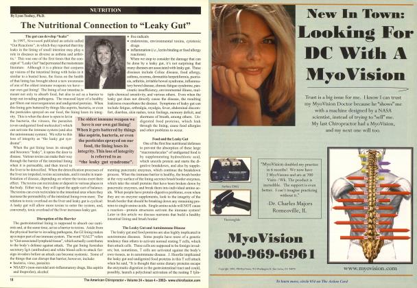
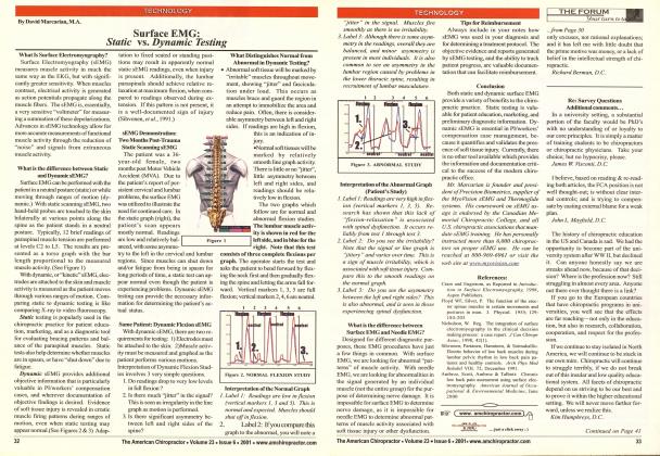
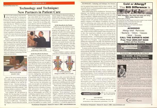
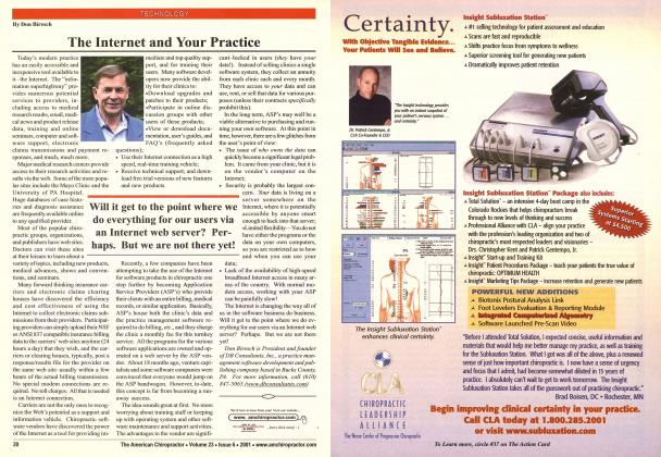
One of the greatest practice challenges for both medical and chiropractic professionals working in the med-legal area is the validation of injury or functional impairment in patients whose objective exam findings do not correlate with their reported symptomatology. Establishing a patient's entitlement to benefits, or even justifying continuing care, can be difficult, when standard ortho/ neuro testing is inconclusive or apparently negative, especially when it is accompanied by a subjective presentation of significant pain or disability. The entry of more objective data into the patient's file can be extremely valuable in establishing the validity of a reported injury, continued impairment, the degree of improvement, ordeeline in the patient's condition, and in helping direct other clinical decisions. And, additional objective findings can directly influence the outcome of personal injury and workers' compensation cases, and other med-legal litigation, as demonstrated here. The following informal case study illustrates how the integration of surface electromyo-graphy (sEMG) and thermography testing can provide the objective findings that standard orthopedic and neurological exam protocols may not elicit. I believe you will concur that these new advances in technology offer doctors the opportunity to obtain critical objective data that has been elusive to clinicians in the past. Details of the Case The patient, in this case, was a 28-year-old male with a history of significant low back pain and radicular symptoms, with an initial onset at eight-years-of-age. The patient had received a prior medical diagnosis, in 1987, of spina bifuda occulta, spondylolisthesis, spondylolysis, and general instability of the lumbosacral junction. Surgical fusion of L5/S1 was recommended and later performed, in September of 1992. Despite several medical opinions that the surgery was successful, the patient reported little improvement in his subjective complaints, when compared pre- and post-surgically. The patient sought intermittent chiropractic care for his persistent complaints during the four-year period between 1995 and 1999. While the patient derived temporary relief from chiropractic care, there was no substantial change in his condition over time. The patient's complaints included significant low back and right leg pain on walking or standing more than three-to-five minutes, and the need to spend an average of 16 hours a day in a non-weight-bearing position to mitigate the pain. The severity of his condition caused him to discontinue employment and seek social security compensation. Several subse- quent medical evaluations yielded insufficient objective findings to support the patient's complaints, and each resulted in denial of a permanent disability rating. The recurring objection was that the patient's subjective complaints and reported level of impairment were not consistent with the results of standard ortho/neuro testing. These inconsistencies contributed to the consideration of a possible psychological origin for the patient's complaints. The patient eventually sought the advice of an attorney, and the treating chiropractor was asked to assist with the identification of information that could support the patient's case. As a result of the numerous medical evaluations, eighteen exhibits had been compiled suggesting that there were insufficient clinical findings to establish permanent disability. In an attempt to find substantiating objective data supporting the patient's subjective complaints, the patient was evaluated utilizing surface EMG and ther-mography systems, in November 1999. A series of detailed computerized reports and graphics were generated. Additionally, the patient received software-based range-of-motion testing, and a complete chiropractic radiological review of all prior films was conducted. The additional objective data obtained, along with the assistance of the patient's legal counsel, were sufficient to contradict the prior eighteen exhibits. This resulted in a legal decision that included a $50,000 award for five years of previously denied benefits, and a determination of permanent disability status, qualifying the patient for social security compensation. The objective data presented was considered to be instrumental in obtaining the favorable legal outcome for this patient. Analysis of the Actual Test Results A. Ihennography Interpretation Fig- I The thermography evaluation was performed twice while the patient maintained a neutral standing posture, with a 15-minute interval between tests (pre- and post-interval.) Note that the red lines indicate the patient's pre- test readings, while the blue lines indicate the post-test results. Temperature readings for the left and right sides of the spine are displayed on the corresponding sides of the graphic. The centerline indicates the differential reading, or the difference between the left and right side temperature readings. In general, the readings should be relatively even when comparing the left and right sides. You can see from the graph that the temperature differential is quite pronounced in the cervical, mid-thoracic, and lumbar regions on the right. What is unusual about this study is the change that occurs upon the second test, after the 15-minute interval. You can clearly see in the circled areas marked #1 that the temperature readings drop significantly in both the mid-thoracic and lumbar areas bilaterally. This may indicate that blood flow was decreased to these regions due to the stress of standing. B. StalicJSurfaceEMG Results Pre- and Post- View— Fig. 2 The static sEMG was also perfomied twice at 15-minute intervals. The patient maintained a neutral standing posture throughout this phase of testing. On the graphic, two sets of horizontal bar graphs appear for each spinal level illustrated. The top bar indicates the Pretest reading, while the lower bar shows the Post-test reading. With the static sEMG test, muscle activity is measured through the application of two probes placed momentarily on the paraspinal areas adjacent to the vertebral levels indicated. This measures the level of muscle tension present at the corresponding spinal segments. Note that, in the Pre- and Post-views, the distance the bars extend laterally from the spine indicates the degree of muscle tension detected. Red indicates a high reading, while purple and green represent moderate and low readings. Under normal circumstances, paraspinal muscles fire evenly, bilaterally, and at relatively low levels of measured activity. Where dysfunction is present, muscles will tense up and "brace" to compensate for the underlying problem. The pattern and level of muscle activity shown in the graphic reveals the muscle bracing exhibited by the patient. When two static sEMG tests are performed within 15 minutes of each other, there should be little change in muscle tension readings, if no intervening adjustment or therapy was performed. It is clear from the area marked #2 that muscle tension increased significantly in the mid-thoracic region on the second test, specifically at T5 and T7 on the right. This may be an indication that the patient was having difficulty standing due to pain, resulting in a compensatory increase in right thoracic muscle tension. C. StatlcJSucfaceJEMG Balance View—Fig. 3 In a normal individual, muscle activity should be relatively balanced between the left and right sides of the spine. The Balance View graphic, presents the Sur- face EMG information in a form that allows us to sec the patient's pattern of bracing. As indicated in this view, there is a clear imbalance in the patient's utilization of paraspinal musculature that correlates with some type of spinal dysfunction. The pattern of muscle activity the patient demonstrates is clearly abnormal and is non-random. The patient demonstrates consistently high activity at the left cervical spine, the right thoracic spine, and the left lumbar spine regions. This is most likely attributable to muscle bracing, which either reduces his pain, or compensates for an underlying structural problem. Dynamic Surface EMG —Fig. 4 This study measures the muscle activity of the lumbar paraspinal regions bilaterally as the patient performs a series of three lumbar flexions. Normally, muscle readings should be relatively low in active flexion, and the muscles should relax at both the full flexion position (markers 1, 3, 5) and at the neutral standing position (markers 2,4,6). Test Observations—Fig. 5 1. Evidence that the test was performed properly is shown by the consistency of the readings on repeated trials. Correct electrode attachment was verified and the patient was provided specific instructions. 2. The lumbar musculature readings reveal "irritability", which is consistent with pain or soft tissue injury. Muscle irritability is demonstrated by thejagged line features, or "jitter", that contrast with the smoother lines of the normal pattern. 3. The muscles do not relax in flexion (markers 1, 3, 5), which is a common finding in patients ex- perieneing pain. 4. Note that there is significant asym- metry in muscle activity, with muscles firing more on the right side than the left. 5. Note that the last repetition of flexion (markers 5 and 6) shows an increase in the irritability of the lumbar paraspinal muscles. This is demonstrated by the amount of "jitter" or perturbations shown on the line graph. This increased variability in the readings may have occurred due to an increase in pain as the third trial began. A patient experiencing pain will show a corresponding increase in the variability of the readings as the number of trials also increases. Test Summary All three of the above tests provide results which are consistent with those typically found in individuals that are experiencing significant pain. The increase in muscle activity and decrease in skin temperature over the 15-minute standing interval also indicate that the patient may have difficulty maintaining an upright posture. Dynamic sEMG shows significant irritability of the muscle groups of the lumbar spine, which is also consistent with patients experiencing pain. Results of these tests should be correlated with the patient's history and all other exam findings. Conclusion This study illustrates the value of additional objective data in substantiating injury and impairment. It is believed that the additional information derived through sEMG and thermog-raphy testing, and the graphic analysis offered by these evaluative methods, had considerable impact on the outcome of this case. This is especially significant considering the volume of medical records and exhibits that were successfully refuted by the inclusion of these test results. David Marcarian, is founder and president of Precision Biometrics, supplier of the MyoVision sEMG and Thermoglide systems. He lectures for Palmer College of Chiropractic, and his course is endorsed by all U.S. chiropractic associations that mandate sEMG training. He has personally instructed more than 6,000 chiropractors on proper sEMG use. He can be reached at 800-969-6961, or visit his company's website at www.mvovision.com.