Solutions for Low Back Pain and Instability
FEATURE
George B. Roth
BSc, DC, ND, CMRP
Low back pain is often associated with lumbar instability. This may be a cause of mechanical stress and inflammation of the intervertebral discs, leading to degeneration, herniation, and chronic pain. Certain injuries appear to initiate a reflex response, whereby the intrinsic paraspinal muscles of the lower lumbar spine become inhibited, leading to articular instability. By determining the precise location of these injuries and treating them using a gentle procedure called Matrix Repatterning, practitioners have been able to restore stability to the lumbar spine, allowing for normalized joint function, reduction of inflammation, and accelerated tissue repair.
Key Terms: Matrix Repatterning, multifidus, rotatores, coupled motion, conus medullaris, cauda equina, sacrificial joints, core injuries, articular stability reflex (ASR), primary restriction.
Throughout the almost 40 years I have been in practice, one of the questions I have always asked myself when confronted with a clinical presentation is why? For example, why do so many people have low back pain? One of my revelations in almost all cases of lumbosacral pain and dysfunction is a significant instability of the lower portion of lumbar spine. This is demonstrable through the application of pressure to the paraspinal sulcus, in which are housed the rotatores and the multifidi. These muscles typically have a significant amount of tone throughout most of the spine1’ 2. However, in the lower lumbar region, it will become evident that the tone is often significantly reduced. In addition, pressure in this region will often elicit localized pain, which is another hallmark of instability3.
The recognition that lumbar instability is a major contributor to low back pain is highlighted by the vast array of devices designed to internally and externally provide artificial stability. Some of these devices, such as lumbar support belts, have been used for hundreds, if not thousands of years.
“For example, why do so many people have low back pain? One of my revelations in almost all cases of lumbosacral pain and dysfunction is a significant instability of the lower portion of lumbar spine. JJ
The idea that any region of the spine would become unstable seems almost counterintuitive, since a major role of the spine is to provide protection for one of the most vital portions of the nervous system, namely the spinal cord. There are no structures that are more heavily protected than the brain and
spinal cord. The spinal cord is particularly vulnerable to mechanical deformation because it occupies almost the entire volume of the spinal canal. This leaves very little tolerance to anything more than a minute amount of intervertebral, segmental joint play. It has been postulated that the paraspinal
muscles perform the role of “neuromuscular ligaments” in the spine, as they allow for a dynamic response to moment-tomoment changes in posture and motion4. This, combined with the articular characteristics of the vertebral column, ensures intervertebral “coupled motion,” which minimizes the possibility for spinal cord compression. The need to safeguard the spinal cord is similar to the protection of communication lines in a military campaign. Without communication, an army would be vulnerable to defeat, and likewise, without a viable neurological communication system, the organism would be vulnerable to injury and death.
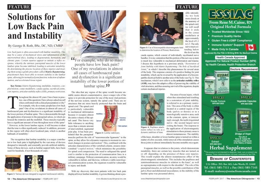
With my discovery that most patients with low back pain displayed lower lumbar instability, it got me thinking about a pos-
sible rationale. As we look more closely at the anatomy of the spinal cord, we will recall that it ends in the conus medullaris at approximately the level of LI to L3. The only
nerve ^ ac^s ex" tending below this level are the
cauda equina, which consist of individually myelinated nerve bundles. It is my contention that this portion of the nervous system is much less vulnerable to mechanical deformation and trauma.
I discuss this hypothesis in a previous article, “Restoration of Joint Stability with Matrix Repatterning: The Role of Sacrificial Joints,” in which I describe this phenomenon for several areas of the body. This response consists of a positive finding for joint bistability, which can be reversed by the application of a biocompatible electrical field to another area of the body (see Fig 3). This mechanism, which I now refer to as the articular stability reflex (ASR), may have the adaptive effect of preserving the viability of vital structures, and thus ensuring survival of the organism, despite certain mechanical injuries.
The area of tissue injury, which when thus stimulated and resulting in a restoration of joint stability, is referred to as a primary restriction. This area of the body is often located in a central region of the body (pelvis or thorax) or in a neurologically sensitive area, such as the cranium, spine, or interestingly enough, the teeth (trigeminal nerve—the second largest nerve exiting the brain). The response to the application of electromagnetic stimulation to these primary areas is almost instantaneous. The stability
of the knee, shoulder or lower lumbar spine is turned on within a matter of a few seconds, while removal of the stimulation causes those joints to almost immediately become unstable once again.
It appears that in relation to the joints, which demonstrate instability, there are certain key muscles that are turned on or off, depending on the presence of these core injuries. This would explain the almost instantaneous effect of the electromagnetic stimulation. This includes the popliteus and semimembranosus muscles in relation to the knee5, as well as supraspinatus with respect to the shoulder6. The role of the paraspinal muscles, as well as the possible contribution of the pelvic floor and abdominal musculature, in the stability of the lumbar spine was presented above.
The guiding principle appeals to be that when there is an injury to certain areas of the body, there is an attempt to mitigate further mechanical damage by turning off stabilizing mechanisms in certain key joints located just peripheral to the trunk of the body. This appears to create a cushioning zone around the core of the body, to increase joint play and thus dissipate or dampen additional mechanical stress, which might potentially damage these vital structures. For example, certain activities, such as gait (lower extremity) and manipulation of loads (upper extremity), have the potential to transmit additional mechanical strain thr oughout the body via fascial and molecular continuity (tensegrity matrix). During gait, the forces transmitted to the pelvis and trunk, upon encountering unusual or irregular surfaces, might be dampened by an unstable knee. In addition, pulling or lifting objects with the upper extremity has the po-
tential to increase mechanical loading of the upper quadrant, including the rib cage and thoracic spine. An unstable shoulder might act to mitigate these forces.
We can theorize that the ASR mechanism is mediated by a spinal reflex, however there is no direct evidence of a specific neural pathway at this time. Nevertheless, the clinical evidence appears to be compelling and has been verified by the observations of a number of practitioners, including physical therapists, chiropractors, and other health professionals, while using these protocols. Our clinical experience, using the Matrix Repatterning program7, has been positive in terms of the correction of primary restrictions in certain core structures and the resultant reestablishment of stability to the lumbar region. In my opinion, joint stability—its causes and effects—are potentially significant contributors to the pain and degenerative changes observed in the lumbar spine. As clinicians, it is imperative that we do our utmost to identify and mitigate these effects to help our patients overcome this painful and debilitating condition.
References:
Fibre type characteristics and function of the human paraspinal muscles: normal values and changes in association with low back pain, Mannion AF, J Electromyog. Kinesiol. Dec 1999, 9 (6): 363-77.
Atrophy of sacrospinal muscle groups in patients with chronic, diffusely radiating lumbar back pain, Laasonen EM, Neuroradiology. 1984:26:9-13.
A New Evaluation Method for Lumbar Spinal Instability: Passive Lumbar Extension Test, Kasai Y, Morishita K, Kawakita E, Kondo T, UchidaA, JAPTA, December 2006.
Lumbar instability: an evolving and challenging concept, Beazell JR, Mullins M, Grindstaff TL, J ManMcmip Diet: 2010Mar; 18 (1): 9-14. Electromyographic Study of the Popliteus Muscle in the Dynamic Stabilization of the Posterolateral Corner Structures of the Knee, K Schinhan, et al, Am J Sports Med, Vol. 39, no. 1, 173-179, January, 2011.
Function of the Supraspinatus Muscle and its Relation to the Supraspinatus syndrome—An Experimental Study in Man, B VanLinge, JDMulder, Journal of Bone and Joint Surgery Vol 45B, No. 4, November 1963, pp 750-754.
The Matrix Repatterning Program for Pain Relief, GB Roth, New Harbinger Publications, Oakland, CA, 2005.
Dr. George Roth has been investigating spinal and musculoskeletal dysfunction for almost 40 years. He is the developer of Matrix Repatterning, a revolutionary technique for the assessment
and treatment of many structural conditions. He is the author of The Matrix Repatterning Program for Pain Relief and the Director of Education for the Matrix Institute. For inquiries, please contact Dr Roth at: g. rathcfmatrixinstitute. net