Applied Kinesiology was first discovered in 1964 by Dr. George Goodheart when he observed that a patient with a winged scapula had a “weak” or inhibited serratus anticus muscle. When Dr. Goodheart applied a technique to refacilitate or “strengthen” the serratus anticus the scapula winging resolved. He eventually coined the term Applied Kinesiology for the application of manual muscle testing as a clinical analyses tool. Dr. Goodheart referred to Applied Kinesiology (AK) or manual muscle testing as functional neurological assessment. Skip forward twenty years to 1984, Dr. Wally Schmitt, one of the most prolific teachers of AK of all time, published a paper describing AK as “Functional Neurology,” or manual muscle testing, as a somatic window into neurological function.
This quote sums up the potential of manual muscle testing, “With few exceptions, all activities of the CNS, receiving, processing, and integrating information, ultimately finds expression in contraction of a muscle,” Color Atlas of Physiology, A. Despopoulos, S. Silberange, Yearbook Medical Publishers, Georg Thieme Verlag. In other words, manual muscle testing can be of value in evaluating nearly every neurological response, reflex, or function in the human system.
Much of the data-gathering process unique to AK relies on manual assessment of muscular function to evaluate changes in the functional status of the nervous system, which are reflected as changes in motor function. These observed changes in muscular function are assumed to be associated with changes in the central integrative state (CIS) of the ventral horn motor neurons.
The CIS is defined as the summation of all excitatory and inhibitory inputs to a neuron.
The functional status or CIS of the ventral horn motor neurons are maintained by convergence of multiple segmental and supra segmental pathways. Segmental pathways are sensory pathways of somatic or visceral origin that arise from receptors in skin, joints, fasciae, viscera, and various chemoreceptors. Supra-segmental pathways are descending pathways of conscious or reflexogenic origin (brainstem, cerebellum) including posturaland gait-pattern generators.
Assuming the alpha motor neuron and associated muscle are free of pathology, a manual muscle test measures the central integrative state of the ventral horn. If the ventral horn CIS is shifted toward hyperpolarization because of inadequate afferent input the alpha motor neuron and associated muscle will reflect this as inhibition. Neuroanatomist Sherrington described the ventral horn-alpha motor neuron and associated muscle as, “the final common pathway.”
Since 2014, when Kevin Tracy, PhD described his research on the vagus nerve and the Cholinergic Anti-inflammatory Pathway, the vagus nerve has permeated health science literature. Here, three different aspects of vagal function, their effects on health and wellness as well as their treatment protocols are discussed, which includevagal autonomic function, Cholinergic Anti-inflammatory function, and vagal immune modulation.
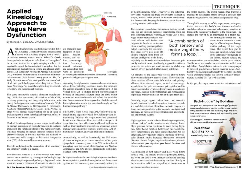
Anatomically, as well as functionally, the 10th cranial nerve or vagus nerve is over one-third of the parasympathetic nervous system. It is 20% motor-efferent projecting from the Dorsal Motor Nucleus and Nucleus Ambiguous, and 80% afferent projecting to the Solitary Nucleus in the brain stem.
In higher vertebrate the two biological systems that learn from experience to defend an organism are the nervous system and the immune system, commonly referred to as the inflammatory reflex. Discovery of the inflammatory reflex revealed that these two systems intersect in simple, precise, reflex circuits to maintain immunological homeostasis, keeping the immune system from becoming overor underactive.
"A combination of both sensory and motor vagal fibers forms a loop."...
The vagus nerve modulates the immune system, including, the gut-immune response, microbiome-brain-gut axis, the innate immune response, as well as CD4 T-cells. Vagal motor output includes visceral efferent cell bodies that originate in the dorsal motor nucleus providing parasympathetic output, especially the intestines.
The vagus nerve also gives off visceral efferent fibers from the nucleus ambiguous to the heart, especially the SA node, which modulates heart rate, primarily to slow it down. And finally, vagus efferent fibers project to the spleen and small intestine to inhibit production of very powerful inflammatory cytokines.
All branches of the vagus with visceral efferent fibers also contain afferent or sensory fibers. The solitary nucleus receives afferent taste information and primary afferents from visceral organs, as well as messenger signals from the gut’s microbiota, gut peptides, and lipopolysaccharides. Cytokines from viscera also activate the vagus, causing the hypothalamus and hippocampus to produce brain cytokines as part of the gut-brain axis.
Generally, vagal signals reduce heart rate, constrict bronchi, increase bronchial secretions, increase peristalsis, modulate intestinal blood flow, activate enzyme release, increase secretions in the stomach, intestines, and pancreas, as well as decrease inflammation and modulate the immune system.
High vagal tone results in better blood-sugar regulation, reduced risk of stroke, cardiovascular disease, lower blood pressure, better resistance to stress, better digestion, better bowel function, better heart rate variability, lower inflammation, and better immune function. On the other hand, low vagal tone leads to increased cardiovascular disease, stroke, increased depression, increased diabetes, chronic fatigue, cognitive impairment, chronic inflammation, poor digestion, poor bowel function, and chronic inflammation.
A combination of both sensory and motor vagal fibers forms a loop. The sensory arc senses pathogens, toxins, and even the body’s own immune molecules sending alerts about excessive inflammatory reactions directly to the brain stem, which are relayed by an interneuron to the motor neurons. The motor neurons then transmit a message to the affected organs through a different path from the vagus nerve, which then completes the loop.
Through the sensory arc of the vagus nerve, pathogens, toxins, and even the body’s own immune molecules send alerts about an excessive inflammatory reaction up through the vagus nerve directly to the brain stem. The signals are relayed by an interneuron to a motor neuron forming the motor arc. The motor neuron transmits a message to affected organs through another pathway of the vagus nerve. This signal then goes to the celiac ganglion, where fibers of the splenic nerve originate. In the spleen, neurons release the neurotransmitter norepinephrine, which prods nearby T-cells to secrete another neurotransmitter called acetylcholine. Acetylcholine interacts with macrophages to diminish production of tumor necrosis factor. Additionally, the vagus projects directly to the small intestine with a cholinergic signal that inhibits the highly inflammatory cytokine Thl7 as well as others.
In the gut, the vagus nerve reads the microbiome and initiates an inflammatory response based on detection of pathologic vs. nonpathologic bacteria. The vagus also simultaneously reads the gut microbiome projecting signals back to the hypothalamus influencing mood, behavior, stress levels, and inflammation.
The vagus feedback loop is considered by many scientists to be the basis for the phrase “gut feeling.” Essentially, this phrase is applicable when one’s body is measuring the state of health and well-being before embarking on a challenging physical or mental activity. In other words, something tells you that today is not the day for this or, “I have a bad feeling about this.”
The cholinergic anti-inflammatory pathway as described by Kevin Tracy, MD, PhD is a neural mechanism that is controlled by the vagus nerve inhibiting local cytokine release from the small intestine and spleen preventing the damaging effect of cytokine over production. In fact, the cholinergic anti-inflammatory pathway is so powerful that vagal implants are now used to successfully treat advanced cases of rheumatoid arthritis.
Researchers were led to believe that the vagus is part of a reflex circuit with a corresponding motor signal referring to organs outside the brain to regulate the inflammatory process.
This vagus immune reflex is the result of inflammatory molecules in tissues, going up the vagus nerve to the brain then returning via the vagus to the original tissues directing them to turn off TNFa as well as any other inflammatory cytokines that might be present.
Stephen Porges, PhD, in his well-received book The Polyvagal Theory describes two separate motor pathways. One is referred to as the ventral vagal complex associated to the nucleus ambiguous and the dorsal vagal complex associated to the dorsal motor nucleus. The parasympathetic effect of the vagus nerve of these two systems will come into play when addressed.
The vagus nerve is known as a cholinergic nerve, or in other words, the neurotransmitter primary to vagal function is acetylcholine. Acetylcholine is synthesized from choline, B-5, B-l, magnesium, manganese, E, B-l2, folic acid, and utilizes zinc at the receptor site. Acetylcholine is diminished by oxidative stress, insulin resistance, mitochondrial dysfunction, high cortisol, insufficient cofactors, brain injury, and reproductive hormone imbalance.
The gold standard for measuring vagal tone is heart rate variability (HRV). Heart rate variability is the time between the R waves in an ECG also referred to as the R-R interval. Greater variability within this interval indicates healthier heart and vagus function. Athletes, especially endurance athletes will have greater HRV than non-exercisers. However, excessive exercise or overtraining will decrease HRV and is an indicator of over exercising.
After this brief review of vagus nerve neurophysiology, it is apparent that nearly every condition that a patient presents with vagus nerve dysfunction should be considered. Whether it is a swollen knee, appetite control, digestive disorders, leaky gut, brain fog, or immune disorders, vagus nerve function should be addressed. Otherwise, the patient can easily fall into a vicious cycle of continual treatment and relapse.
Treatment of vagus nerve dysfunction is much easier than the physiology implies. There are many products that are currently marketed as vagus nerve stimulatory devices. These devices can be quite effective under the right circumstances. However, if there is an underlying physiological or structural issue, the results are temporary at best. Therefore, it is best to address chemical and structural imbalances before applying stimulation devices and exercises.
Upper cervical spine fixation and subluxations are commonly associated to vagus nerve dysfunction. C1-C3 segments project afferents to the nucleus intermedius then to the nucleus tractus solitarius (NTS) of the vagus nerve. If there is upper cervical fixation or subluxation normal mechanoreceptor afferentation to the NTS is lost with consequent vagus dysfunction. Additionally, the temporal bulge cranial fault as described by Walther et al., is commonly associated to vagus nerve dysfunction and digestive disorders. And finally, the acupuncture points GB1 are associated to acetylcholine function.
The simple approach to vagal dysfunction is to locate an inhibited muscle associated to the area of pain, inflammation, digestive dysfunction, or immune system dysfunction, or any other malady. Utilizing the involved muscle therapy localize (TL) the vagus point just inside the sternal notch. If TL to the vagal point facilitates the involved muscle it implies vagal involvement. Depending on what condition is being addressed applying nociception (pinching) over the visceral referred pain areas associated to the small intestines, spleen, heart, or lungs, will inhibit a normally facilitated (strong) muscle. This inhibited muscle will be used to localize the areas to be treated. Utilizing the inhibited muscle, TL the upper cervical spine, GB 1 acupuncture points, and over the side of the cranium above the ear. Make the appropriate corrections as indicated. Then address the involved chemical faults or nutrition, apply electrical stimulation or TENS to the points in the ear as well as activation exercises to achieve long lasting and health-changing correction.
Correcting Cholinergic Anti-inflammatory Pathway
1. Vagal point in sternal notch TL facilitates an inhibited muscle
a. Pinching spleen and/or small intestine VRPs inhibit globally
b. Treat individual VRPs with temporal bulge cranial fault, upper cervical segmental correction, and GB1 stimulation
Correcting Vagal Immune and Autonomic Modulation
1. Vagal point TL in sternal notch facilitates visceral related muscles
a. Pinching heart and/or lung VRPs inhibits globally i. Ventral vagal complex/nucleus ambiguous
dysfunction
b. Pinching digestive organ VRPs inhibits globally
i. Dorsal vagal complex/dorsal motor nucleus dysfunction
c. Treat individual VRP’s with temporal bulge cranial fault correction, upper cervical segmental correction, and GB 1 stimulation
Correction Exercises for Vagal Dysfunction
• Gargling, rub abdomen, abdominal pressure, humming, or repeating sound “OM” (Vagus nerve innervates vocal cords)
• Slow rhythmic diaphragmatic breathing
• Meditation, especially loving kindness meditation
Normally they have the patient perform one of these activities three times a day for as many weeks as it takes to correct dysfunction
Electrical Stimulation Points
Dr. Belli the current president of the International College of Applied Kinesiology graduated magna cum laude from Life Chiropractic College West in 1984. Dr. Belli is a Fellow of the American Board of Neurochemistry and Nutrition and a Diplomat of the American Chiropractic Association Council on Neurology. Additionally, Dr. Belli has spent the last 30 years researching and developing advanced systems of neurologically based Applied Kinesiology. You may contact Dr. Belli [email protected].