As chiropractors, we are well aware of how a weak core can impact a patient’s ability to hold a chiropractic adjustment. The strength of the abdominal and pelvic floor muscles, especially the transverse abdominis (TA), plays a crucial role in maintaining spinal and pelvic alignment. However, for most of the population, inactivity, poor posture, or incorrect exercises have caused these muscles to be deconditioned, and the rectus abdominis muscles are separated at the midline, called a diastasis recti. Although diastasis recti is very common because of pregnancy, nonpregnant adults and even children can also suffer from diastasis recti with symptoms including back pain, incontinence, digestive issues like constipation or bloating, and umbilical hernia. It is important to check every patient for diastasis recti if they are complaining of any of the previously mentioned symptoms.
Checking for Diastasis Recti
Professionals should look for two things when they check for a diastasis recti — the distance between the separated muscles and the condition of the connective tissue.
Most professionals check the distance of the diastasis at its smallest instead of at its largest because when they look for a diastasis, they ask the client to lift their head and shoulders. When the head and shoulders come off the floor, the muscles come closer together, and it is then examined at its smallest. Before assessing the distance of the diastasis, it is important to see if the person has a doming of their abdominal muscles when they lift their head or if they have an umbilical hernia. The presence of either or both conditions indicates that they need to be checked in a different manner. If they do have doming or a hernia, then the distance is evaluated on each side of the umbilicus. If they do not have it, then the distance is checked in the middle of the belly. Either way, the diastasis is examined in three places. It is checked at the umbilicus, above the umbilicus (halfway between the sternum and the umbilicus), and below the umbilicus (halfway between the umbilicus and the pubic bone).
Looking for a diastasis is done in a back-lying position with the knees bent. If checking on each side of the belly for a large diastasis, you use eight closed fingers with the fingers pointing toward the toes. The distance is evaluated after having them then relax their abdominal muscles and engage them by bringing their umbilicus to the spine. When they bring the umbilicus to the spine, the muscles will come closer together, and you will feel the ridges of the muscles. When they are relaxed again, you follow the muscles to where they go in a relaxed position. In this relaxed position, you are measuring the separation at its largest. The muscles are engaged and relaxed several times so you can feel the movement of the muscles.
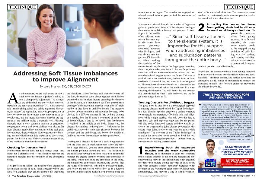
..." Since soft tissue attaches to the skeletal system, it is imperative for this support when addressing imbalances and subluxation patterns throughout the entire body....”
You do each side and then add the number of fingers together to get the total distance. If there is not a doming of the muscles or umbilical hernia, then you put 10 closed fingers in the middle of the belly and measure it the same way in the same three places previously mentioned. You start with 10 fingers, and can always take fingers out if it is smaller. When checking the condition of the connective tissue, the deeper the finger goes down into the tissue, the weaker that tissue is. Put the finger in the umbilicus with the abdominal muscles relaxed, and then see where the skin goes against the finger. This can be marked with a pen on the finger; shallow is up to 2 cm, moderate is around 4 cm, and deep is 6 cm or greater. The condition of connective tissue is checked in the same places above and below the umbilicus, like when checking the distance. You will know that the connective tissue is healing when it gets shallower, and the finger does not go down as far.
Treating Diastasis Recti Without Surgery
The good news is that there is a nonsurgical approach to treating diastasis recti called the Tupler Technique® . It is four simple steps that empower a patient to restore core strength, and therefore, effectively hold their alignment while weight bearing. Not only does this lead to less back pain and improved digestion, but the patient will also notice improved posture and theoretically decrease the degenerative joint disease progression that occurs when joints are receiving repetitive stress while misaligned. The outcome of the Tupler Technique® is to make the linea alba strong enough to hold the recti muscles in a close together position. It utilizes three key components in healing diastasis recti.
1. Repositioning both the separated muscles and the weak connective tissue. It is important to align the separated muscles close together so that both the muscles and connective tissue move in the sagittal plane when engaging the transverse muscle during activities of daily living and when doing the Tupler Technique® exercises. When the muscles are four fingers apart or more without being approximated, they move in a side-to-side direction instead of front-to-back direction. The connective tissue needs to be continuously put in a narrow position to take the stretch off it and allow it to heal.
2. Protecting the connective tissue from getting stretched in either a forward or sideways direction. To
protect the connective tissue from getting stretched in a forward direction, the transverse muscle needs to be engaged during activities of daily living, exercise routines, and sporting activities. Also, exercises done in a downward-facing abdominal position should be avoided.
To prevent the connective tissue from getting stretched in a sideways direction, avoid activities where the back is arched. This flares the ribs, and besides stretching the connective tissue, makes it impossible to engage the transverse muscle. The forward crossover movement should also be avoided.
3. Strengthening the abdominal muscles and connective tissue with the Tupler Technique® exercises. Research on connective tissue by Dr. Helene M. Langevin, a neurologist at the University of Vermont, formulated a medical hypothesis suggesting that connective tissue might comprise an elaborate communication system within the body, which influences the function of all the other physiological systems. In her article, “Connective Tissue: A Body-Wide Signaling Network?” she explores the possibility that there might be a series of remote effects in seemingly unrelated parts of the body stemming from activity in the connective tissue. She also talks about the connective tissue generating electric currents through mechanical activity, including compression, which could change the cellular activity in any given interconnected parts of the body. Hypothetically, the compression in the specific Tupler Technique® exercises creates a microcurrent in the connective tissue, and that remodels and heals the connective tissue.
Proper Alignment While Weight Bearing
We all have seen those patients who are challenged when it comes to holding their chiropractic adjustments. They feel amazing when they get off the table, and after a couple of days, they slowly return to their old subluxated self. When joints are unable to “stay in place” after being aligned, we need to investigate the root cause(s).
In addition to addressing a weak core by knitting together a diastasis recti, patients also need to be checked for collapse of one or more arches of the feet. Like splinting the abdominal muscles to support the correct position of the soft tissue in a patient with diastasis recti, custom-made orthotics that provide three-arch support remind the fascia and connective tissue of where they are optimal. Since soft tissue attaches to the skeletal system, it is imperative for this support when addressing imbalances and subluxation patterns throughout the entire body. After all, it is all connected.
Laura Brayton, DC, CSP, CSCP, CACCP, is a graduate of New York Chiropractic College and the I University of North Carolina at Chapel Hill. As a holistic chiropractor and speaker, she holds certifications in Chiropractic Pediatrics, Webster Technique for breech presentation, Sacro-Occipital Technique (S.O.T.), Craniopathy, is an advanced level practitioner of Nambudripad's Allergy Elimination Technique (NAET) and a Tupler Technique® trainer.
She is the owner and founder of Hoboken Family Chiropractic + Wellness, in Hoboken, NJ. Follow her on FB and IG @drlaurabrayton and at www.drlaurabrayton.com.