Like the character Neo in the movie The Matrix, I decided to take the red pill many years ago by questioning the concepts that were being taught in school and the various seminars I attended over the years.
Eventually, my skepticism paid off, and I was fortunate enough to discover certain principles that have been verified by reproducible scientific evidence and the clinical experience of practitioners and patients around the world.
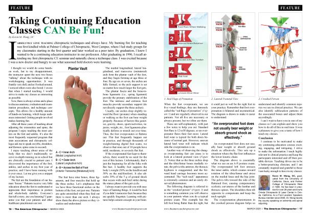
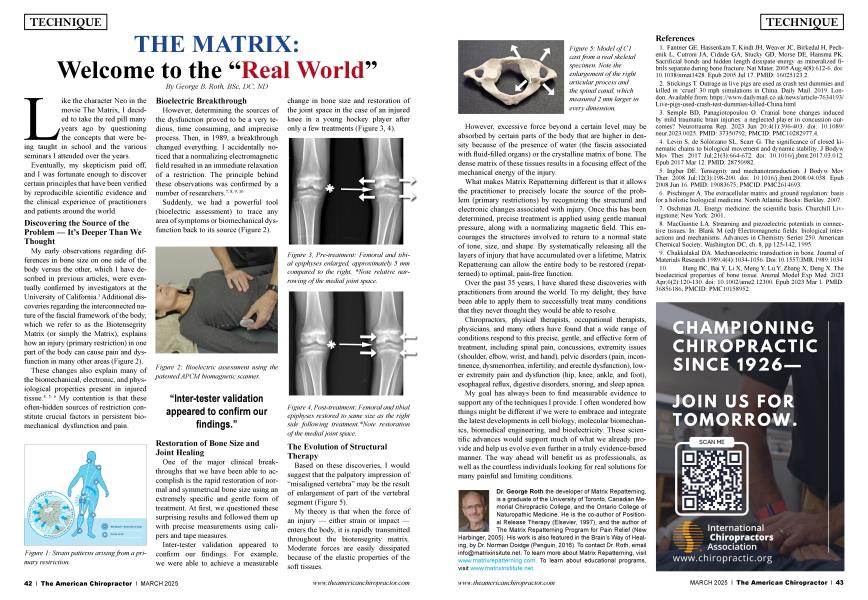
My early observations regarding differences in bone size on one side of the body versus the other, which I have described in previous articles, were eventually confirmed by investigators at the University of California.1 Additional discoveries regarding the interconnected nature of the fascial framework of the body, which we refer to as the Biotensegrity Matrix (or simply the Matrix), explains how an injury (primary restriction) in one part of the body can cause pain and dysfunction in many other areas (Figure 2).
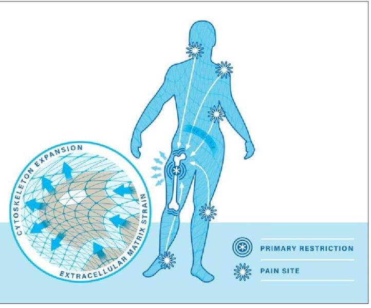 Figure 1: Strain patterns arising from a primary restriction.
These changes also explain many of the biomechanical, electronic, and physiological properties present in injured tissue.41516 My contention is that these often-hidden sources of restriction constitute crucial factors in persistent biomechanical dysfunction and pain.
However, determining the sources of the dysfunction proved to be a very tedious, time consuming, and imprecise process. Then, in 1989, a breakthrough changed everything. I accidentally noticed that a normalizing electromagnetic field resulted in an immediate relaxation of a restriction. The principle behind these observations was confirmed by a number of researchers.7 819110
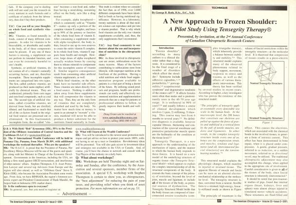
Suddenly, we had a powerful tool (bioelectric assessment) to trace any area of symptoms or biomechanical dysfunction back to its source (Figure 2).
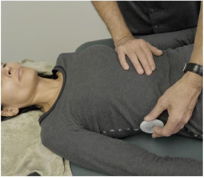 Figure 2: Bioelectric assessment using the patented APCM biomagnetic scanner.
“Inter-tester validation appeared to confirm our findings.”
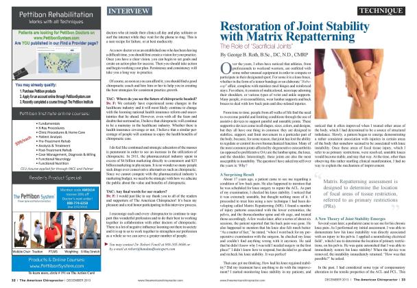
One of the major clinical breakthroughs that we have been able to accomplish is the rapid restoration of normal and symmetrical bone size using an extremely specific and gentle form of treatment. At first, we questioned these surprising results and followed them up with precise measurements using calipers and tape measures.
Inter-tester validation appeared to confirm our findings. For example, we were able to achieve a measurable change in bone size and restoration of the joint space in the case of an injured knee in a young hockey player after only a few treatments (Figure 3, 4).
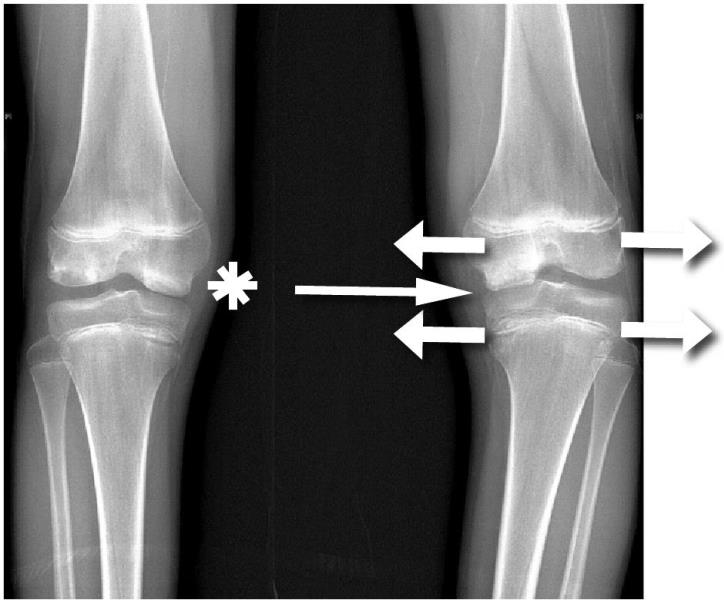 Figure 3, Pre-treatment: Femoral and tibial epiphyses enlarged, approximately 5 mm compared to the right. *Note relative narrowing of the medial joint space.
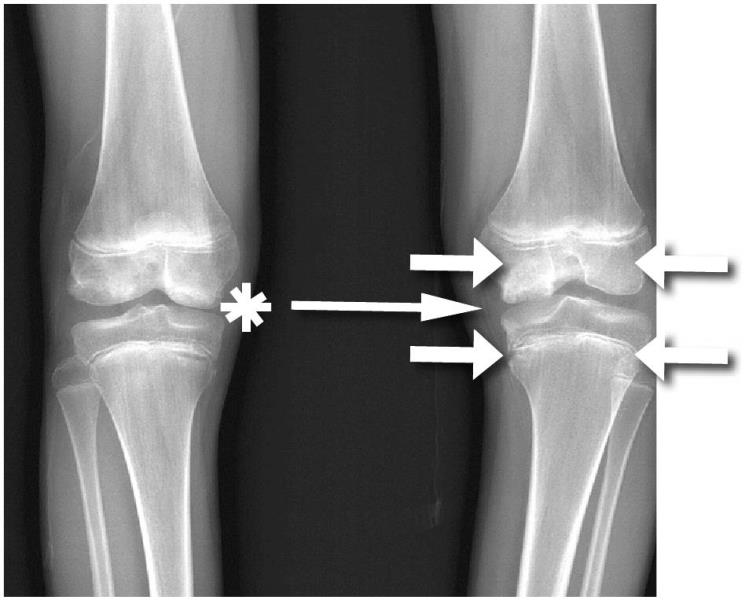 Figure 4, Post-treatment: Femoral and tibial epiphyses restored to same size as the right side following treatment. *Note restoration of the medial joint space.
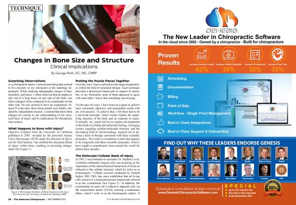
Based on these discoveries, I would suggest that the palpatory impression of “misaligned vertebra” may be the result of enlargement of part of the vertebral segment (Figure 5).
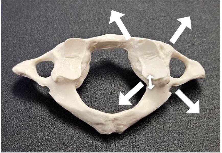 Figure 5: Model of C1 cast from a real skeletal specimen. Note the enlargement of the right articular process and the spinal canal, which measured 2 mm larger in every dimension.
My theory is that when the force of an injury — either strain or impact — enters the body, it is rapidly transmitted throughout the biotensegrity matrix. Moderate forces are easily dissipated because of the elastic properties of the soft tissues.
However, excessive force beyond a certain level may be absorbed by certain parts of the body that are higher in density because of the presence of water (the fascia associated with fluid-filled organs) or the crystalline matrix of bone. The dense matrix of these tissues results in a focusing effect of the mechanical energy of the injury.
What makes Matrix Repatterning different is that it allows the practitioner to precisely locate the source of the problem (primary restrictions) by recognizing the structural and electronic changes associated with injury. Once this has been determined, precise treatment is applied using gentle manual pressure, along with a normalizing magnetic field. This encourages the structures involved to return to a normal state of tone, size, and shape. By systematically releasing all the layers of injury that have accumulated over a lifetime, Matrix Repatterning can allow the entire body to be restored (repattemed) to optimal, pain-free function.
Over the past 35 years, I have shared these discoveries with practitioners from around the world. To my delight, they have been able to apply them to successfully treat many conditions that they never thought they would be able to resolve.
Chiropractors, physical therapists, occupational therapists, physicians, and many others have found that a wide range of conditions respond to this precise, gentle, and effective form of treatment, including spinal pain, concussions, extremity issues (shoulder, elbow, wrist, and hand), pelvic disorders (pain, incontinence, dysmenorrhea, infertility, and erectile dysfunction), lower extremity pain and dysfunction (hip, knee, ankle, and foot), esophageal reflux, digestive disorders, snoring, and sleep apnea.
My goal has always been to find measurable evidence to support any of the techniques I provide. I often wondered how things might be different if we were to embrace and integrate the latest developments in cell biology, molecular biomechanics, biomedical engineering, and bioelectricity. These scientific advances would support much of what we already provide and help us evolve even further in a truly evidence-based manner. The way ahead will benefit us as professionals, as well as the countless individuals looking for real solutions for many painful and limiting conditions.
Dr. George Roth the developer of Matrix Repatterning, is a graduate of the University of Toronto, Canadian Memorial Chiropractic College, and the Ontario College of Naturopathic Medicine. He is the co-author of Positional Release Therapy (Elsevier, 1997), and the author of The Matrix Repatterning Program for Pain Relief (New Harbinger, 2005). His work is also featured in the Brain’s Way of Healing, by Dr. Norman Doidge (Penguin, 2016). To contact Dr. Roth, email [email protected]. To learn more about Matrix Repatterning, visit www.matrixrepatterning.com. To learn about educational programs, visit www.matrixinstitute.net.
References
1. Fantner GE, Hassenkam T, Kindt JH, Weaver JC, Birkedal H, Pechenik L, Cutroni JA, Cidade GA, Stucky GD, Morse DE, Hansma PK. Sacrificial bonds and hidden length dissipate energy as mineralized fibrils separate during bone fracture. Nat Mater. 2005 Aug;4(8):612-6. doi: 10.1038/nmatl428. Epub 2005 Jul 17. PMID: 16025123.2.
2. Stickings T. Outrage as live pigs are used as crash test dummies and killed in 'cruel' 30 mph simulations in China. Daily Mail. 2019. London. Available from: https://www.dailymail.co.uk/ne... Live-pigs-used-crash-test-dummies-killed-China.html
3. Semple BD, Panagiotopoulou O. Cranial bone changes induced by mild traumatic brain injuries: a neglected player in concussion outcomes? Neurotrauma Rep. 2023 Jun 20;4(l):396-403. doi: 10.1089/ neur.2023.0025. PMID: 37350792; PMCID: PMC10282977.4.
4. Levin S, de Solorzano SL, Scarr G. The significance of closed kinematic chains to biological movement and dynamic stability. J Bodyw Mov Ther. 2017 Jul;21(3):664-672. doi: 10.1016/j.jbmt.2017.03.012. Epub 2017 Mar 12. PMID: 28750982.
5. Ingber DE. Tensegrity and mechanotransduction. J Bodyw Mov Ther. 2008 Jul; 12(3): 198-200. doi: 10.1016/j.jbmt.2008.04.038. Epub 2008 Jun 16. PMID: 19083675; PMCID: PMC2614693.
6. Pischinger A. The extracellular matrix and ground regulation: basis for a holistic biological medicine. North Atlantic Books: Berkley. 2007.
7. Oschman JL. Energy medicine: the scientific basis. Churchill Livingstone: New York. 2001.
8. MacGuintie LA. Streaming and piezoelectric potentials in connective tissues. In: Blank M (ed) Electromagnetic fields: biological interactions and mechanisms. Advances in Chemistry Series 250. American Chemical Society, Washington DC, ch. 8, pp 125-142, 1995.
9. Chakkalakal DA. Mechanoelectric transduction in bone. Journal of Materials Research. 1989;4(4): 1034-1056. Doi: 10.1557/JMR. 1989.1034
10. Heng BC, Bai Y, Li X, Meng Y, Lu Y, Zhang X, Deng X. The bioelectrical properties of bone tissue. Animal Model Exp Med. 2023 Apr;6(2): 120-130. doi: 10.1002/ame2.12300. Epub 2023 Mar 1. PMID: 36856186; PMCID: PMC10158952.