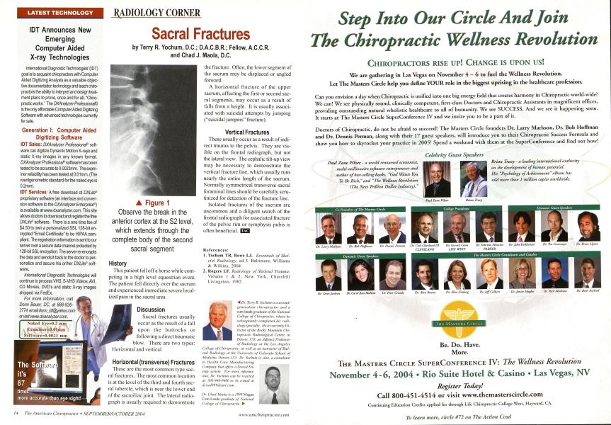
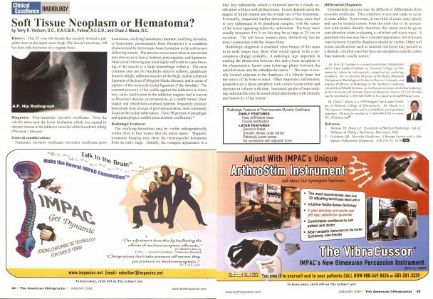
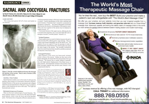
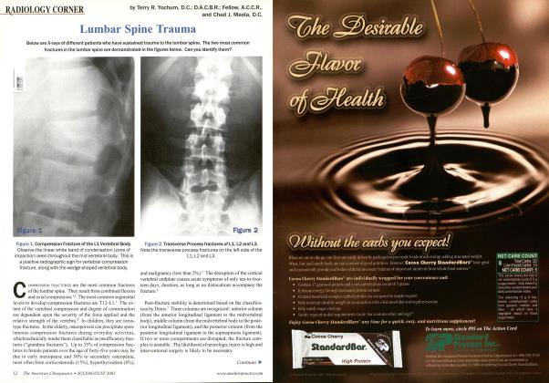
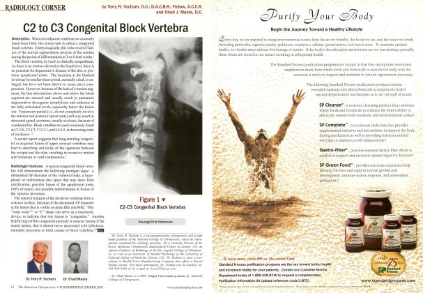
IDT Announces New Emerging Computer Aided X-ray Technologies International Diagnostic Technologies' (IDT) goal is to acquaint chiropractors with Computer Aided Digitizing Analysis as a valuable objective documentation technology and teach chiropractors the ability to interpret and design treatment plans to prove, once and for all, "Chiropractic works." The DXAnalyzer Professional® is the only affordable Computer Aided Digitizing Software with advanced technologies currently for sale. Generation I: Computer Aided Digitizing Software IDT Sales: DXAnalyzer Professional' software can digitize Dynamic Motion X-rays and static X-ray images in any known format. DXAnalyzer Professional software has been tested to be accurate to 0.0023mm. The examiner reliability has been tested at 0.01 mm. (The roentgenometric standard for the naked eye is 0.2mm). IDT Services: A free download of DXLite® proprietary software (an interface and conversion software to the DXAnalyzer Enterprise®) is available atwww.dxanalyzer.com. This site allows doctors to download and register the free DXLite' software. There is a one time fee of $4.50 to own a personalized SSL 128-bit encrypted "Email Certificate" to be HIPAA compliant. The registration information is sent to our server over a secure data channel protected by 128-bit SSL encryption. The server re-encrypts the data and sends it back to the doctor to personalize and secure his or/her DXLite' software. International Diagnostic Technologies will continue to process VHS, S-VHS Videos, AVI, CD Movies, DVD's and static X-ray images shipped via FedEx. For more information, call Donn Bauer, DC, at 888-926-2774, email [email protected] History This patient fell off a horse while competing in a high level equestrian event. The patient fell directly over the sacrum and experienced immediate severe localized pain in the sacral area. Discussion Sacral fractures usually occur as the result of a fall upon the buttocks or following a direct traumatic blow. There are two types: Horizontal and vertical. Horizontal (transverse) Fractures These are the most common type sacral fractures. The most common location is at the level of the third and fourth sacral tubercle, which is near the lower end of the sacroiliac joint. The lateral radiograph is usually required to demonstrate the fracture. Often, the lower segment of the sacrum may be displaced or angled forward. A horizontal fracture of the upper sacrum, affecting the first or second sacral segments, may occur as a result of falls from a height. It is usually associated with suicidal attempts by jumping ("suicidal jumpers" fracture). Vertical Fractures These usually occur as a result of indirect trauma to the pelvis. They are visible on the frontal radiograph, but not the lateral view. The cephalic tilt-up view may be necessary to demonstrate the vertical fracture line, which usually runs nearly the entire length of the sacrum. Normally symmetrical transverse sacral foraminal lines should be carefully scrutinized for detection of the fracture line. Isolated fractures of the sacrum are uncommon and a diligent search of the frontal radiograph for associated fracture of the pelvic rim or symphysis pubis is often beneficial. EZS References: Yochum TR, Rowc LJ. Essentials of Skel etal Radiology, ed 3. Baltimore. Williams & Wilkins, 2004. Rogers LF. Radiology of Skeletal Trauma, Volume I & 2, New York. Churchill Livingston. 1982. ■^ Dr. Terry R. Yochwn is a second-generation chiropractor and a cum laude graduate of the National College of Chiropractic, where he subsequently completed his radiology specialty. He is currently Director of the Rocky Mountain Chiropractic Radiological Center, in Denver. CO. an Adjunct Professor oj Radiology at the Los Angeles College of Chiropractic, as well as an instructor of Skeletal Radiology at the University of Colorado School of Medicine, Denver, CO. Dr. Yoclmm is. also, a consultant to Health Care Manufacturing Company that offers a Stored Energy system. For more information. Dr. Yochum can he reached at: 303-940-9400 or hy e-mail at dcradO99(a aal.com. Dr. Chad Maola is a 1999 Mayna Cum Laude graduate of National College of Chiropractic. ► A Figure 1 Observe the break in the anterior cortex at the S2 level, which extends through the complete body of the second sacral segment