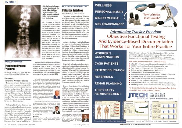
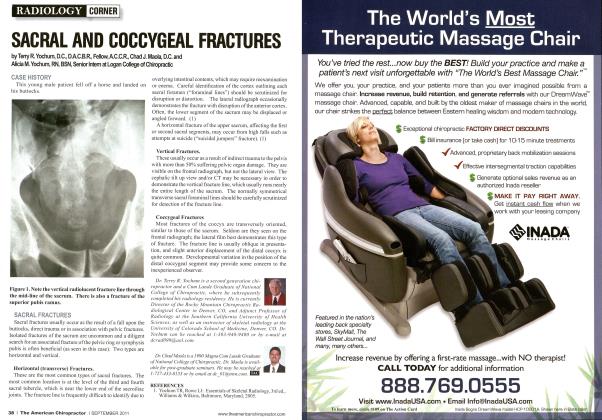
Discussion Transverse Process Fractures Transverse process fractures arc the second most common fractures of the lumbar spine, with compression fractures being the most common. They occur usually secondary to a severe hyperextension and lateral flexion blow to the lumbar spine from avulsion of the paraspinal muscles. The most common segments to suffer transverse process fractures are L2 and L3. (Figure above) Radiographically, the fracture line appears as a jagged radiolucent separation, usually occurring close to its point of origin from the vertebra. Frequently, the separated fragment is displaced infe-riorly. If the fracture line is horizontal, close inspection for a transverse or Chance fracture should be performed. Fractures often occur at multiple lev- els. Fractures of the fifth lumbar transverse process are frequently found in association with pelvic fractures, particularly fractures of the sacral ala, or disruption of the sacroiliac joint. Occasionally, loss of the psoas shadow may occur secondary to hemorrhage. Ossification within this hemorrhage (myositis os-sificans) can result in bony bridging between transverse processes (lumbar ossified bridging syndrome). Renal damage may occur, which may be associated with hematuria. A pseudofraeture of the transverse process can be simulated by developmental nonunion, especially at LI, the psoas margin where it crosses the tip of the transverse process and overlying fat lines or intestinal gas. Oblique or tilt views may be necessary to rule out fracture. IB3 Dr. Teny R. Yochum is a second generation chiropractor and a Cum Laudc Graduate of the National College of Chiropractic, where he subsequently completed his radiology residency. He is currently Director of the Rocky Mountain Chiropractic Radiological Center in Denver, Colorado, and Adjunct Professor of Radiology at the Southern California University of Health Sciences, as well as an instructor of skeletal radiology at the University of Colorado School of Medicine, Denver, CO. Dr. Yochums 3'J edition textbook. Essentials of Skeletal Radiology, was released in the fall of2004 and is now available for purchase. Dr. Yochum can be reached at 303-940-9400 or by e-mail at dcrad099(a)aol.corn. Dr. ChadJ. Maola is a 1990 Magna Cum Laude Graduate of the National College of Chiropractic. Dr. Maola has co-authored five chapters in Dr. Yochum .V 3"1 edition textbook and is rendering post-graduate lectures with Dr. Yochum and separately throughout the United States. Dr. Maola is a Chiropractic Orthopedist ami is available for post-graduate seminars. He may be reached at 303-690-8503 or e-mail DC_0 [email protected]. Reference Yochum TR, Rowc LJ, Essentials of Skeletal Ra-iliology. 3"1 ed. Lippincott, Williams and Wilkins, Baltimore, 2005. Note the irregular fracture vertical lines through the left L1,L2 and L3 transverse processes. The callus formation adjacent to the fracture suggests they are healing.