Case Presentation
RADIOLOGY
Cliff Tao
DC, DACBR
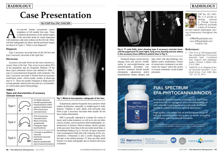
A 41-year-old female recreational runner complains of left medial foot pain. There is bilateral prominence at the medial aspect of the navicular tarsal on visual inspection with tenderness and some redness on the left side. Bilateral foot radiographs are taken, and the pertinent images are shown in Figure 1. What is your diagnosis?
Diagnosis
Type 2 accessory navicular bone of the left foot and type 3 accessory navicular of the right foot.
Discussion
Accessory navicular bones are the most common accessory bone in the foot. They occur in up to about 20% of the population and are frequently bilateral. Of the three types commonly known and outlined in Table 1, type 2 is associated most frequently with symptoms. The type 2 accessory navicular is formed from an accessory ossification center, which usually ossifies at about age nine to 11. These are usually triangular in shape and are at least partially united to the navicular by a synchondrosis with hyaline and/or fibrocartilage.
TABLE 1.
Types and characteristics of accessory navicular bones.
Symptomatic patients frequently have posterior tibial tendon dysfunction, especially in middle-aged or older patients. Children or early adults with navicular pain from a type 2 accessory navicular bone usually do not have tendon abnormality.
MRI is generally indicated to evaluate for extent of injury and to plan treatment, as well as to rule out other sources of pain, such as posterior tibial tendinopathy, osteonecrosis, or fracture. Marrow edema, which is greater in the accessory bone than in the navicular tarsal bone, is the hallmark finding (Fig 2). Severity of injury increases with synchondrosis fluid and with widening of the synchondrosis. Ultrasound is also a useful imaging tool, but only in the skilled operator. CT does not offer much benefit over plain radiographs due to limited soft-tissue evaluation.
Treatment begins conservatively, ranging from just activity modification to non-weight-bearing cast immobilization. Secondary conservative treatment should include chiropractic adjustments and/or manipulations, dietary changes, and pain relief with physiotherapy modalities and/or medications. Failure of conservative treatment is an indication for surgery where the ossicle is excised, commonly via the Kidner procedure.
Cliff Tao, DC, DACBR, is in private radiology practice in Orange County, California, and leads a team of chiropractic and medical radiologists, serving chiropractors throughout the world.
www.clifftaodcdacbr.com
714-876-1126
References:
1. Choi YS et al. MR imaging findings of painful type II accessory navicular bone: Correlation with surgical and pathologic studies, Korean J Radiol 5(4): 274-9, 2004.
2. Tuthill HL et al. Imaging of tarsal navicular disorders: A pictorial review. Foot Ankle Spec 7(3): 211-25, 2014.