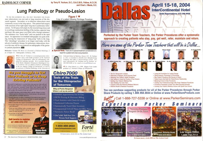
IN THE I'KE-ANIIBIOIIf ERA, THE ONLY TREATMENT FOR PULMO- nary tuberculosis was rest and or lung resection of the diseased segment. Secondary or re-infection tuberculosis most commonly occurs in the lung apices. This patient had surgery with resection of her right upper lung due to pulmonary tuberculosis. To fill space left empty by the resected lung tissue and to avoid huge mediastinal shifting of the trachea, opposite lung and heart, this open space was tilled with a foreign substance. The substance was "lucite balls" and was packed in the open space. Its appearance on standard radiographs was quite striking, leaving the impression of "ping pong" balls in the lung. This "plombage" procedure is no longer used today with the advent of antibiotic therapy for pulmonary tuberculosis; however this may still be encountered on radiographs of the geriatric patient population. EZS Reference: Yochum TR. Roue LJ; Essentials of Skeletal Radiology. 2 ed. (Chapter 16 - Radiographic Artifacts), 1996. ^ Dr. Terry R. Yochum is a second-general ion chiropractor and a cum laude graduate of the National College of Chiropractic, where he subsequently completed his radiology specialty. He is currently Director of the Rocky Mountain Chiropractic Radiological Center, in Denver. CO. an Adjunct Professor of Radiology ut the Los Angeles College of Chiropractic, as well as an instructor of Skeletal Radiolog}' at the University of Colorado School of Medicine, Denver, CO. Dr. Yochtun is, also, a consultant to Health Care Manufacturing Company that offers a Stored Energy system. For more information. Dr. Yochum can be reached at: 303-940-9400 or hy e-mail at daad099(<r(iol,com. ■^ Dr. Chad Maola is a 1999 Magna Cum Laude graduate of National College of Chiropractic. ▲ Special Note: There are no special treatment considerations to report for a patient who had a lung resection. Figure 1 X-ray of a patient following Plombage Procedure