By George B. Roth, DC, ND, CMRP
The first time I visited the Canadian Memorial Chiropractic College in Toronto, even before enrolling in 1974, I decided to check out the dissection lab. I met a senior student there who was employed as a lab assistant. He graciously described the specimen he was working on in detail.
I was enthralled by the amazing and beautiful complexity of the inner workings of the human body. I was hooked!
I decided to enroll in the program and learn everything I could about anatomy. I realized that the best way to do that would be to teach it, which led me to eventually become an anatomy lab assistant.
Teaching dissection for approximately three years gave me the opportunity to learn the intricate details of anatomy in great depth. One thing I noticed was a commonly occurring variation in the size and shape of the bones on one side of the body versus the other side.
I made similar observations in radiology classes. In those days (1970s in Ontario), if one knee, hip, or shoulder was being X-rayed, it was mandatory that the same structure on the opposite side also be radiographed. This important tool is no longer routinely used but could be very helpful in determining the significance of a structural finding.
When I mentioned these variations to the anatomy and radiology professors, they simply shrugged it off as “normal variations.” At the time, I didn’t realize the implications and simply filed them away.
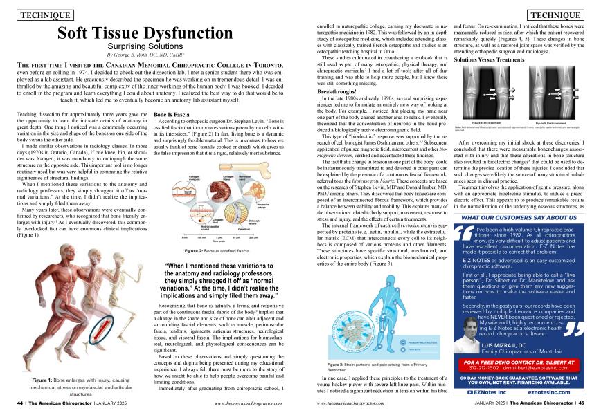
Many years later, these observations were eventually confirmed by researchers, who recognized that bone literally enlarges with injury.1 As I eventually discovered, this commonly overlooked fact can have enormous clinical implications (Figure 1).
According to orthopedic surgeon Dr. Stephen Levin, “Bone is ossified fascia that incorporates various parenchyma cells within its interstices.” (Figure 2) In fact, living bone is a dynamic and surprisingly flexible material. This is in contrast to how we usually think of bone (usually cooked or dried), which gives us the false impression that it is a rigid, relatively inert substance.
Recognizing that bone is actually a living and responsive part of the continuous fascial fabric of the body,2 implies that a change in the shape and size of bone can alter adjacent and surrounding fascial elements, such as muscle, perimuscular fascia, tendons, ligaments, articular structures, neurological tissue, and visceral fascia. The implications for biomechanical, neurological, and physiological consequences can be significant.
Based on these observations and simply questioning the concepts (and dogma) being presented during my educational experience, I always felt there must be more to the story of how we might be able to help people overcome painful and limiting conditions.
Immediately after graduating from chiropractic school, I enrolled in naturopathic college, earning my doctorate in naturopathic medicine in 1982. This was followed by an in-depth study of osteopathic medicine, which included attending classes with classically trained French osteopaths and studies at an osteopathic teaching hospital in Ohio every month for five years.
These studies culminated in coauthoring a textbook that is still used as part of many osteopathic, physical therapy, and chiropractic curricula.3 I had a lot of tools after all of that training and was able to help more people, but I knew there was still something missing.
In the late 1980s and early 1990s, several surprising experiences led me to formulate an entirely new way of looking at the body. In 1989, I noticed that placing my hand near one part of the body caused another area to relax. I eventually theorized that the concentration of neurons in the hand produced a biologically active electromagnetic field.
That “bioelectric” response was supported by the research of cell biologist James Oschman and others.4,5 Subsequent application of pulsed magnetic fields and biomagnetic microcurrent generators verified and accentuated these findings.
The fact that a change in tension in one part of the body could be instantaneously transmitted to and detected in other parts can be explained by the presence of a continuous fascial framework, referred to as the biotensegrity matrix. These concepts are based on the research of Stephen Levin, MD6 and Donald Ingber, MD, PhD,7 among others. They discovered that body tissues are composed of an interconnected fibrous framework, which provides a balance between stability and mobility. This explains many of the observations related to body support, movement, response to stress and injury, and the effects of certain treatments.
The internal framework of each cell (cytoskeleton) is supported by proteins (e.g., actin, tubulin), while the extracellular matrix (ECM) that interconnects every cell to its neighbors is composed of various proteins and other filaments. These structures have specific structural, mechanical, and electronic properties, which explain the biomechanical properties of the entire body (Figure 3).
In 1990, as I applied gentle pressure on the enlarged tibial plateau of a patient with knee pain, I felt a sudden change in the tension within the bone. On reexamination, I noticed that the bone was measurably reduced in size. Following the treatment, the patient recovered remarkably quickly (Figures 4, 5).
After overcoming my shock at these discoveries, I speculated that there were objective bone changes associated with injury, and these changes also produced measurable bioelectric changes that could be used to assess the precise location of injury. Such changes were likely the source of many structural imbalances seen in clinical practice.
By applying gentle pressure, along with an appropriate bioelectric stimulus to induce a piezoelectric effect, it is possible to produce remarkable results in the normalization of the underlying osseous structures, as well as the surrounding and adjacent soft tissues.8 The fact that these results last and are permanent in many cases promises to provide real solutions, as opposed to ongoing and often endless treatments.9,10
These discoveries and the emerging science of the structure of the body at the deepest levels have helped explain many of my clinical and biomechanical observations and how these properties provide us with the opportunity to help our patients at the most profound level.
The technique that ultimately emerged from these discoveries is called Matrix Repatterning, and it has been applied by practitioners from all over the world. It has helped tens of thousands of individuals suffering from a myriad of conditions, such as back, neck, and shoulder pain; carpal tunnel syndrome; hip, knee, and ankle pain; TMJ (temporomandibular joint) dysfunction; headaches; and concussions.11 Matrix Repatterning has also helped patients with other functional conditions, such as esophageal reflux, snoring, sleep apnea, cardiovascular issues, digestive disorders, urinary incontinence, and erectile dysfunction, and the list keeps growing.
About the Author
Dr. George Roth is a graduate of the University of Toronto, Canadian Memorial Chiropractic College, and the Ontario College of Naturopathic Medicine. He also has studied osteopathic medicine at Doctors' Hospital North in Columbus, Ohio. Dr. Roth is the developer of Matrix Repatterning and the director of education at the Matrix Institute in Toronto. He has presented seminars at numerous hospital and university-based symposia throughout North America. He is the coauthor with Kerry D’Ambrogio, PT, of Positional Release Therapy (Elsevier, 1997) and the author of The Matrix Repatterning Program for Pain Relief (New Harbinger, 2005). His work is also featured in The Brain’s Way of Healing by Dr. Norman Doidge (Penguin, 2016).
To learn more about Matrix Repatterning, visit www.matrixrepatterning.com. Learn more about educational programs at www.matrixinstitute.net.
References:
Fantner GE, Hassenkam T, Kindt JH, Weaver JC, Birkedal H, Pechenik L, Cutroni JA, Cidade GA, Stucky GD, Morse DE, Hansma PK. Sacrificial bonds and hidden length dissipate energy as mineralized fibrils separate during bone fracture. Nat Mater. 2005 Aug;4(8):612-6. Epub 2005 Jul 17.
Blottner D, Huang Y, Trautmann G, Sun L. The fascia: continuum linking bone and myofascial bag for global and local body movement control on Earth and in space. a scoping review. REACH. 2019;14–15. doi:10.1016/j.reach.2019.100030.
Roth GB, D’Ambrogio K. Positional release therapy. New York: Elsevier Science; 1997.
Oschman JL. Energy medicine: the scientific basis. New York: Churchill Livingstone; 2001.
MacGuintie LA. Streaming and piezoelectric potentials in connective tissues, In: Blank M (ed). Electromagnetic fields: biological interactions and mechanisms. Advances in Chemistry Series 250. Washington DC: American Chemical Society.; 1995;125-142.
Levin S. The importance of soft tissues for structural support of the body. Spine: State of the Art Reviews. 1995: 9(2).
Ingber DE. The architecture of life. Sci Am. 1998 Jan;278(1):48-57. doi: 10.1038/scientificamerican0198-48. PMID: 11536845.
Pischinger A. The extracellular matrix and ground regulation: basis for a holistic biological medicine. Berkley: North Atlantic Books; 2007.
Roth GB. The matrix repatterning program for pain relief. Oakland, CA: New Harbinger Publications; 2005.
Roth GB. Matrix repatterning: advanced structural therapy. Toronto: Matrix Institute; 2019.
Doidge N. The brain’s way of healing. New York: Penguin Books; 2016.