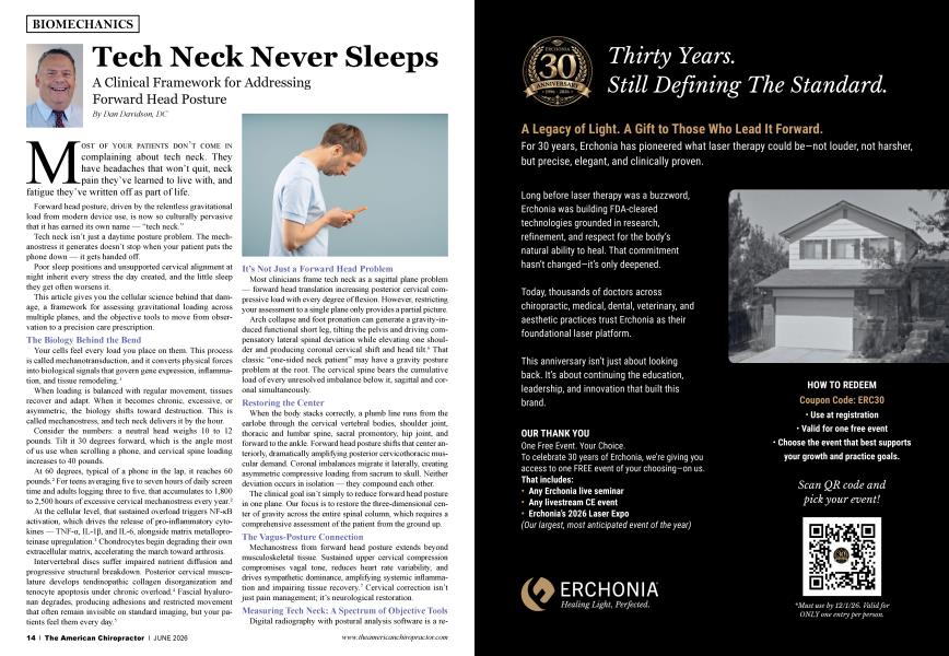
MOST OF YOUR PATIENTS DON’T COME IN complaining about tech neck. They have headaches that won’t quit, neck pain they’ve learned to live with, and fatigue they’ve written off as part of life.
Forward head posture, driven by the relentless gravitational load from modern device use, is now so culturally pervasive that it has earned its own name — “tech neck.”
Tech neck isn’t just a daytime posture problem. The mechanostress it generates doesn’t stop when your patient puts the phone down — it gets handed off.
Poor sleep positions and unsupported cervical alignment at night inherit every stress the day created, and the little sleep they get often worsens it.
This article gives you the cellular science behind that damage, a framework for assessing gravitational loading across multiple planes, and the objective tools to move from observation to a precision care prescription.
Your cells feel every load you place on them. This process is called mechanotransduction, and it converts physical forces into biological signals that govern gene expression, inflammation, and tissue remodeling.1
When loading is balanced with regular movement, tissues recover and adapt. When it becomes chronic, excessive, or asymmetric, the biology shifts toward destruction. This is called mechanostress, and tech neck delivers it by the hour.
Consider the numbers: a neutral head weighs 10 to 12 pounds. Tilt it 30 degrees forward, which is the angle most of us use when scrolling a phone, and cervical spine loading increases to 40 pounds.
At 60 degrees, typical of a phone in the lap, it reaches 60 pounds.2 For teens averaging five to seven hours of daily screen time and adults logging three to five, that accumulates to 1,800 to 2,500 hours of excessive cervical mechanostress every year.2
At the cellular level, that sustained overload triggers NF-KB activation, which drives the release of pro-inflammatory cytokines — TNF-a, IL-10, and IL-6, alongside matrix metalloproteinase upregulation.3 Chondrocytes begin degrading their own extracellular matrix, accelerating the march toward arthrosis.
Intervertebral discs suffer impaired nutrient diffusion and progressive structural breakdown. Posterior cervical musculature develops tendinopathic collagen disorganization and tenocyte apoptosis under chronic overload.4 Fascial hyaluronan degrades, producing adhesions and restricted movement that often remain invisible on standard imaging, but your patients feel them every day.5
Most clinicians frame tech neck as a sagittal plane problem — forward head translation increasing posterior cervical compressive load with every degree of flexion. However, restricting your assessment to a single plane only provides a partial picture.
Arch collapse and foot pronation can generate a gravity-induced functional short leg, tilting the pelvis and driving compensatory lateral spinal deviation while elevating one shoulder and producing coronal cervical shift and head tilt.6 That classic “one-sided neck patient” may have a gravity posture problem at the root. The cervical spine bears the cumulative load of every unresolved imbalance below it, sagittal and coronal simultaneously.
When the body stacks correctly, a plumb line runs from the earlobe through the cervical vertebral bodies, shoulder joint, thoracic and lumbar spine, sacral promontory, hip joint, and forward to the ankle. Forward head posture shifts that center anteriorly, dramatically amplifying posterior cervicothoracic muscular demand. Coronal imbalances migrate it laterally, creating asymmetric compressive loading from sacrum to skull. Neither deviation occurs in isolation — they compound each other.
The clinical goal isn’t simply to reduce forward head posture in one plane. Our focus is to restore the three-dimensional center of gravity across the entire spinal column, which requires a comprehensive assessment of the patient from the ground up.
Mechanostress from forward head posture extends beyond musculoskeletal tissue. Sustained upper cervical compression compromises vagal tone, reduces heart rate variability, and drives sympathetic dominance, amplifying systemic inflammation and impairing tissue recovery.7 Cervical correction isn’t just pain management; it’s neurological restoration.
Digital radiography with postural analysis software is a reliable method for objective cervical assessment. Lateral views allow direct measurement of lordosis angle, anterior head translation, and disc space height. Deviations from the coronal plane, such as lateral listhesis, scoliotic curvature, and vertebral rotation, are visible in AP views.
Analysis software serves as a clinical baseline and template for treatment strategies. Research consistently supports radiographic postural analysis for tracking structural change under chiropractic care.8
Grid-based postural screening applications using standard photography provide a practical entry point that is useful for documenting asymmetries, tracking gross postural change, and generating patient education materials. They can serve as valuable adjuncts, particularly for screening and wellness care.
Structured light and infrared imaging map tens of thousands of surface data points in a single scan, generating three-dimensional postural models. This captures bilateral weight distribution, center of mass position, and asymmetries across both planes simultaneously.
When applied to the cervical region, this technology precisely captures the three-dimensional geometry of the head, neck, and shoulder complex, enabling objective, individualized support recommendations. This area is where measurement and personalized care intersect most powerfully.
We spend nearly one-third of our lives sleeping9 When cervical aligmnent goes unsupported during those hours, the mechanostress your patients accumulate all day doesn’t pause — it compounds. This can block the disc rehydration, tissue recovery, and neurological restoration that sleep should provide.
Tech neck is typically framed as a daytime problem. The mechanostress accumulated across hours of screen time and forward head loading doesn’t clock out at bedtime, though — it continues, depending on the positions your patients sleep in and the support they are probably lacking. Educating patients about sleep position, posture, and support is not a secondary conversation.
Stomach Sleeping: The most mechanically destructive common sleep position for the cervical spine. Prone sleeping forces sustained cervical rotation, creating uneven disc loading, joint compression, foraminal nanowing on the turned side, and chronic posterior muscle strain.10
It also extends the lumbar spine and rotates the thoracic spine, directly undermining the postural corrections you’re working to establish. Transitioning committed stomach sleepers to side or back sleeping is a clinical priority.
Side Sleeping: When properly supported, side lying maintains excellent cervical aligmnent. The problem is the semi-fetal habit with the upper shoulder rolled forward, head tilting toward the mattress, which drives lateral cervical flexion, shoulder compression, and thoracic rotation.
Research confirms that neck pain patients spend significantly more time in provocative sleep postures than asymptomatic controls, and that these are ingrained habits, not comfort positions.11 The critical variable is pillow height; it must precisely fill the shoulder-to-head gap to maintain neutral aligmnent.
Back Sleeping: This is the preferred position for cervical recovery. Supine sleeping distributes weight evenly and preserves the spine’s natural sagittal curves without rotation. Pillow selection is everything — too flat allows cervical extension, but too thick recreates the forward flexion you’re clinically correcting. A retrospective study of 410 cervical pain patients showed significant symptomatic improvement following individualized pillow height adjustment.12
No generic pillow can serve a diverse patient population. Shoulder width, neck length, head size, thoracic kyphosis, and dominant sleep position all interact to determine each patient’s specific cervical support needs.
Research confirms that very low and very high pillow heights produce significantly greater cervical muscle activation than properly fitted individualized support. Optimal height correlates directly with the body’s dimensions.13
Patients using CPAP machines present a particular challenge because standard cervical pillows frequently conflict with mask positioning and hose routing, leading patients to abandon either CPAP compliance or cervical support — both clinical losses.
This is the clinical rationale for individualized cervical support prescribed from objective measurement, which is the same principle driving custom orthotic fabrication, now applied to the nocturnal cervical environment. A truly customized pillow created from the patient’s head, neck, and shoulder data is optimal for both back and side sleeping. Accoimnodating CPAP without the loss of optimal support transforms sleeping hours from a mechanostress liability to a therapeutic asset.
The following protocol integrates everything previously mentioned into a reproducible clinical workflow:
• Identify the full load. Document sagittal and coronal plane imbalances through digital radiography, postural scanning, or functional screening. Assess kinetic chain contributors from the feet up and include nocturnal posture habits during the intake.
• Remove abnormal load first. Custom orthotics correct gravitational imbalances at the foundation. Ergonomic modification and postural retraining reduce daytime mechanostress. Individualized cervical support pillows address the nocturnal third.
• Restore through corrective care. Chiropractic adjustments deliver beneficial mechanotransduction, restoring segmental mobility, activating Piezo mechanoreceptor pathways, and normalizing neural function. 14 Soft-tissue work resolves fascial adhesions. Cervical corrective exercise rebuilds deep flexor strength and scapular stability.
• Measure and document progress. Objective reassessment, cervical lordosis angle, postural scan data, and center-of-gravity position all demonstrate clinical impact in terms that patients can more easily understand.
Tech neck is ancient biology ambushed by modern technology, building mechanostress across every waking and sleeping hour. As chiropractors, we are uniquely positioned to address it throughout the full 24-hour cycle with objective measurement documenting the load and foundational structural correction to redistribute it.
Chiropractic corrective care restores the biology, and custom cervical support protects the sleeping enviromnent where healing should happen. When we apply the complete clinical solution, we are not merely treating a condition. We change a trajectory for optimal future health and function.

Dr. Dan Davidson is a Palmer College of Chiropractic graduate in Davenport, Iowa. He has been in practice for over 40 years and owns The Back Resort & Peak My Health Center in Salem, Virginia. Dr. Davidson also hosts the Peak My Health podcast, sharing tips on health, exercise, nutrition, and posture. He is also a member of the Foot Levelers Speakers Bureau. Learn more at www.footlevelers.com/more.
1. Ingber DE. Mechanobiology and diseases of mechanotransduction. Ann Med. 2003;35(8):564-77. doi: 10.1080/07853890310016333. PMID: 14708967.
2. Hansraj KK. Assessment of stresses in the cervical spine caused by posture and position of the head. Surg Technol Int. 2014 Nov;25:277-9. PMID: 25393825.
3. Guilak F, Fermor B, Keefe FJ, Kraus VB, Olson SA, Pisetsky DS, Setton LA, Weinberg IB. The role of biomechanics and inflammation in cartilage injury and repair. Clin Orthop Relat Res. 2004 Jun;(423): 17-26. doi: 10.1097/01.blo.0000131233.83640.91. PMID: 15232421.
4. Sharma P, Mafiulli N. Tendon injury and tendinopathy: healing and repair. J Bone Joint Surg Am. 2005 Jan;87(l): 187-202. doi: 10.2106/JBJS.D.01850. PMID: 15634833.
5. Stecco C, Stem R, Porzionato A, Macchi V, Masiero S, Stecco A, De Caro R. Hyaluronan within fascia in the etiology of myofascial pain. Surg Radiol Anat. 2011 Dec;33(10):891-6. doi: 10.1007/s00276-011-0876-9. Epub 2011 Oct 2. PMID: 21964857.
6. Rothbart BA, Estabrook L. Excessive pronation: a major biomechanical determinant in the development of chondromalacia and pelvic lists. J Manipulative Physiol Ther. 1988 Oct;ll(5):373-9. PMID: 2976805.
7. Wang H, Gao X, Shi Y, Wu D, Li C, Wang W. Effects of trunk posture on cardiovascular and autonomic nervous systems: a pilot study. Front Physiol. 2022 Oct 18:13:1009806. doi: 10.3389/fphys.2022.1009806. PMID: 36330208; PMCID: PMC9623330.
8. Harrison DD, Janik TJ, Harrison GR, Troyanovich S, Harrison DE, Harrison SO. Chiropractic biophysics technique: a linear algebra approach to posture in chiropractic. J Manipulative Phvsiol Ther. 1996 Oct;19(8):52535. PMID: 8902664.
9. Gordon S, Grimmer K, Trott Patricia. Sleep position, age, gender, sleep quality and waking cervicothoracic symptoms. Internet J Allied Health Set Pract. 2007;5(l). doi: 10.46743/1540-580X/2007.1134
10. Cary D, Jacques A, Briffa K. Examining relationships between sleep posture, waking spinal symptoms and quality of sleep: a cross sectional study. PLoS One. 2021 Nov 30;16(ll):e0260582. doi: 10.1371/journal. pone.0260582. Erratum in: PLoS One. 2024 Jul 2;19(7):e0306662. doi: 10.1371/journal.pone.0306662. PMID: 34847195; PMCID: PMC8631621.
11. Gordon SJ, Grimmer-Somers KA, Trott PH. Pillow use: the behavior of cervical stiffness, headache and scapular/arm pain. J Pain Res. 2010 Aug 11:3:137-45. doi: 10.2147/jpr.sl0880. PMID: 21197317; PMCID: PMC3004642.
12. Yamada S, Hoshi T, Toda M, Tsuge T, Matsudaira K, Oka H. Changes in neck pain and somatic symptoms before and after the adjustment of the pillow height. JPhvs Ther Sci. 2023 Feb;35(2):106-113. doi: 10.1589/ jpts.35.106. Epub 2023'Feb 1. PMID: 36744195; PMCID: PMC9889209.
13. Ren S, Wong DW, Yang H, Zhou Y, Lin J, Zhang M. Effect of pillow height on the biomechanics of the head-neck complex: investigation of the cranio-cervical pressure and cervical spine alignment. Peer J. 2016 Aug 31;4:e2397. doi: 10.7717/peeij.2397. PMID: 27635354; PMCID: PMC5012320.
14. Pickar JG. Neurophysiological effects of spinal manipulation. Spine J. 2002 Sep-Oct;2(5):357-71. doi: 10.1016/sl529-9430(02)00400-x. PMID: 14589467.