THE VERTEBRAL SUBLUXATION COMPLEX, particularly its histophysiopathology, is a contentious issue within the medical and chiropractic communities.
Vertebral subluxation is a term widely used in chiropractic to describe a purported misalignment of the vertebrae believed to affect nerve function and overall health. However, the concept has been met with skepticism by much of the broader medical and scientific community due to a lack of rigorous scientific evidence supporting its existence.
Secondary effects caused by spinal manipulation have been demonstrated through various physiological and central neurological effects, primarily due to its impact on the central nervous system (CNS) and the way it influences pain perception, proprioception, and overall neurological function. Here are some of the key secondary physiological and central neurological effects associated with spinal manipulation:
a. Endorphin release: Spinal manipulation can stimulate the release of endorphins and other neuropeptides, which help modulate pain perception. This can lead to a reduction in pain symptoms for conditions such as headaches, back pain, and musculoskeletal pain.1
b. Gate control theory: The manipulation may activate mechanoreceptors in the skin and muscles, which can inhibit pain signals traveling to the brain, thereby reducing the perception of pain.2
a. Neuroplastic changes: Spinal manipulation may facilitate neuroplastic changes in the central nervous system (CNS), allowing for improved adaptation and recovery from injury. This can be particularly beneficial for chronic pain conditions, where maladaptive neural pathways may contribute to ongoing pain.3
a. Proprioceptive feedback: By improving spinal alignment and joint mobility, spinal manipulation can enhance proprioceptive feedback to the brain. This can contribute to better coordination and balance, as well as improved awareness of body position.4
a. Changes in cortical activity: Studies using neuroimaging techniques have shown that spinal manipulation can affeet cortical areas of the brain involved in pain processing and motor control. This may lead to changes in how pain is perceived and how movement is coordinated.5 6
a. Sympathetic and parasympathetic balance: Spinal manipulation may influence the autonomic nervous system by promoting a balance between sympathetic (fight or flight) and parasympathetic (rest and digest) responses. This can lead to reductions in stress and anxiety, which can also affect pain perception and overall well-being.7
a. Reduction in central sensitization: In conditions characterized by central sensitization (where the CNS becomes hypersensitive to stimuli), spinal manipulation may help reduce this sensitivity and improve pain outcomes.8
All these secondary changes occur because of spinal manipulation at any nonspecific spinal level.
The vertebral subluxation complex is a primary cellular, tissue, and anatomical lesion altering neuronal transmission quality and quantity attributable to cell mechanics. Neuronal mechanotransduction has been extensively studied in sensory neurons that transduce mechanical stimuli related to hearing, sensation, and pain.
Physical forces on sensory nerve endings open various ion channels, including mechanosensitive ion channels and receptors. Recent research indicates that mechanosensitive ion channels are widely present in various cells and tissues, suggesting that mechanosensitivity is also prevalent in the central nervous system. This would make the vertebral subluxation the primary origin of altered vitalistic function.9
The absence of a universally accepted definition of a vertebral subluxation complex makes it challenging to conduct research that can be widely accepted. There is a need for basic, valid scientific theory with an understanding of the underlying cellular/ tissue dysfunction of the vertebral subluxation complex.
In my opinion, chiropractic institutions should be spearheading innovative research on the influence of mechanobiology and mechanotransduction as major contributing factors to the basis of the vertebral subluxation complex’s histophysiopathology. Nerve tissue, extracellular matrix, cytoskeleton, and cellular membrane through mechanotransduction could give us a working theory to work upon, and some basic science research already exists.
Displacement of any part of the skeletal frame may press against (or stretch) nerves, which are the channels of communication, intensifying or decreasing their carrying capacity, creating either too much or not enough functioning, an aberration known as “dis-ease.” Chiropractors adjust, by hand, all displacements of the more than 200 bones, specifically those of the vertebral column, to remove nerve impingement or tension that causes deranged function.
Mechanical force is known to affect a diversity of physiological areas at the cellular level in all cells, including nerve, cardiac, fibroblast, bone, and vascular cells. Mechanotransduction is a topic of increasing research interest, particularly to those in the neural sciences, because of the ability of physically based forces to induce neuronal biochemical changes directly responsible for a host of complex and integrated neurological cell responses.
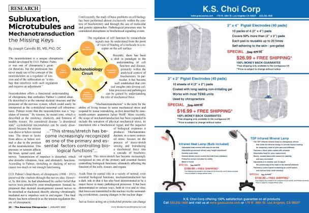
Mechanotransduction, in a more restricted sense, focuses on the process of stress/stretch sensing and transducing a mechanical force into a cascade of biochemical signals. This stress/stretch has become increasingly recognized as a primary and essential factor controlling biological functions, ultimately affecting the function of the cells, tissues, and organs.
All living things, despite their profound diversity, share a common architectural building block: the cell. Cells are the basic functional units of life, even though they are comprised of many components with distinct mechanical characteristics.
Cells control and undergo many intra and extracellular events for their functions. The subject of cell mechanics encompasses a wide range of essential cellular processes, ranging from macroscopic events, such as the maintenance of cell shape, cell motility, adhesion, and deformation, to microscopic events, such as how cells sense mechanical signals and transduce them into a cascade of biochemical signals leading to a host of biological responses.
Cell mechanics recently underwent rapid development and research with particular attention to the rheology of the cytoskeleton and the reconstituted gels of some of the major cytoskeletal components, actin filaments, intermediate filaments, microtubules, and their cross-linking proteins that are responsible for the main structural properties and motilities of the cell. Another area of intense investigation is the mechanical interaction of the cell with its surroundings — cell to cell and extracellular matrix (ECM) — and how this interaction causes changes in cell morphology and biological signaling that lead to functional adaptation or pathological conditions.
On describing the mechanics of living cells, modeling the cytoskeleton as a simple mechanical elastic, viscoelastic, or poor-viscoelastic continuum, a porous gel or soft glassy material non-Newtonian fluid, or a tensegrity (tension integrity) network incorporating discrete structural elements that bear compression.
The importance of biology in cell mechanics is most evident in the ability of the cell to sense and respond to gravity and applied forces. All cells can sense applied physical force. They respond to force through a variety of biological pathways that lead to such diverse consequences as changes in membrane channel activity, upor down-regulation of gene expression, alterations in protein synthesis, or altered cell morphology.
Forces in vivo are often transmitted to the cell via the extracellular matrix, which shares in the load-supporting function. Many cell membrane receptors contain extracellular domains that bind to the various proteins of the extracellular matrix (ECM). For example, members of the integrin family receptors can bind to fibronectin, vitronectin, collagen, and laminin.
Intracellular domains of these same proteins bind directly (or indirectly, through other membrane-associated proteins) to the cytoskeleton. Of these transmembrane molecules (both proteins and proteoglycans), many attach directly to the cytoskeleton, which often exhibits a denser, more rigid structure in the vicinity of an adhesion site.10-11
A thin lipid bilayer, rich with phospholipids, glycolipids, cholesterol, and transmembrane proteins, separates cells from the external environment. Phospholipids are the most abundant and are amphipathic, having a hydrophilic part residing on the outside surface of the bilayer and a hydrophobic part on the inside.
Some of the proteins serve as ion channels, others as a pathway for transmembrane signaling. Still others provide a structural bridge across the membrane, allowing for direct adhesion between the internal cytoskeleton and the extracellular matrix. We refer to these collectively as integral membrane proteins.
Roughly half of the integral proteins freely diffuse, while the rest anchor to the cytoskeleton. In addition to its role in communicating stress and biochemical signals into the cell, the membrane also serves as a barrier function, isolating the cell’s interior from its extracellular environment and maintaining the appropriate biochemical conditions within for critical cell functions. By itself, the bilayer generally contributes little to the overall stiffness of the cell; the microtubule contributes to cellular morphology, communication, and stiffness.12
Microtubules constitute a major constituent of the cytoskeleton. Monomers of aand P-tubulin construct polymerized filaments into a hollow cylinder. The filaments have an outer diameter of about 25 nm and exhibit a high bending stiffness. Tubular structures tend to be more resistant to bending than solid cylinders with the same amount of material per unit length, and that, combined with the larger radius, accounts for the high bending stiffness of microtubules.
Intermediate filaments (IFs) constitute a superfamily of proteins containing more than 50 different members. They have in common a structure consisting of a central a-helical domain of over 300 residues that forms a coiled-coil. Aside from these differences in structure, intermediate filaments differ from microfilaments and microtubules in terms of their long-term stability.13
Of course, these are only a few of the many proteins that contribute to the mechanical properties of a cell. The cytoskeleton primarily consists of actin filaments, microtubules, and intermediate filaments.
In addition to these are the molecular constituents of the cell membrane, nuclear membrane, all the organelles, and other intracellular bodies that influence the overall mechanical response of a cell. While pharmacologic-related research has dominated basic science research on nerve stimulation and function, the effects of mechanical impact on nerve function (mechanotransduction) are receiving increased attention.
Researchers at the Institute of Biomedical Sciences (Taipei, Taiwan) and at Carnegie Mellon University (Pittsburgh, PA) conducted a 2009 study and published findings in an article titled “Understanding Sensory Nerve Mechanotransduction through Localized Elastomeric Matrix Control. ” The researchers used in vitro methods to evaluate mechanical stretch effects on neuromusculoskeletal structures, focusing on the effect of stretch-activated mechanotransduction on nerve terminal action potentials.14
Indentation displacement was applied in increments of 10.43 um until an action-potential (AP) response occurred or displacement reached a maximum of 125 um. When an indentation produced an AP, the same indentation was applied again to see whether the stretch-activated AP was repeatable; it was repeatable virtually 100% of the time.
Displacement lasted less than one second, with a minimum of 30 seconds between indentations. “Specific mechanical deformations and extracellular matrix (ECM) interactions trigger neural AP firing,” concluded the authors.14 These findings have implications in the field of chiropractic, manual medicine, and physical therapy.
Mechanotransduction in neural tissue is the process by which mechanical forces are converted into biochemical signals that can influence the behavior and function of neurons and other cells within the nervous system. Neural tissue is sensitive to mechanical cues and can respond to changes in mechanical forces through various mechanisms.
One of the key components involved in mechanotransduction in neural tissue is the cytoskeleton, which provides structural support and plays a role in cell shape, motility, and intracellular signaling. Changes in mechanical forces can lead to alterations in cytoskeletal organization and dynamics, impacting neuronal morphology and function.
Mechanical forces can also influence ion channels and membrane receptors in neurons, leading to changes in membrane potential and neuronal excitability. For example, stretch-activated ion channels in neuronal membranes can be activated by mechanical forces, altering ion flow and signaling within the cell.
In addition, mechanotransduction in neural tissue can impact cellular processes, such as neurite outgrowth, synaptic plasticity, and gene expression. Mechanical cues from the extracellular enviromnent, such as the stiffness of the surrounding matrix, can influence neuronal development and connectivity.
Overall, mechanotransduction in neural tissue is a complex and dynamic process that plays a critical role in neuronal function, development, and adaptation to the mechanical environment. Understanding how mechanical forces are sensed and transduced by neural cells can provide insights into neural pathophysiology and the potential to understand the chiropractic vertebral subluxation complex.
Mechanotransduction is the process by which cells convert mechanical stimuli into biochemical signals, which can then influence cellular behavior and function. This process is relevant in various physiological and pathological contexts, including the vertebral subluxation complex (VSC).
The vertebral subluxation complex is a term used primarily in chiropractic to describe a condition where a vertebra is misaligned or otherwise not functioning properly relative to the vertebrae above and below it. Misalignment can affect nerves, muscles, and tissue function, leading to various symptoms and dysfunctions.
When a vertebra is misaligned because of trauma, poor posture, inflammation, and visceral-somatic reflexes, it can cause mechanical stress on the surrounding tissues, including muscles, ligaments, and intervertebral disc. It can also induce changes in the extracellular matrix, leading to altered nerve microtubule function transmitting altered nerve function and communication.
A stiff extracellular matrix can limit the range of motion of vertebrae and surrounding tissues, making it difficult to maintain proper spinal alignment and function. The ECM plays a role in the microenvironment surrounding nerve tissues. Stiffening may affect nerve function and communication, leading to pain or dysffinction associated with the vertebral subluxation complex.
Mechanotransduction allows cells in these tissues to sense and respond to these changes. For example:
1. Mechanical response
Cells can sense mechanical changes through mechanoreceptors, which may trigger a cascade of events involving the cytoskeleton, including microtubules. This response can lead to cellular adjustments that aim to maintain homeostasis or adapt to the altered mechanical enviromnent.
a. Fibroblasts in ligaments and tendons may alter their production of collagen and other extracellular matrix components in response to altered mechanical loads, stiffening the extracellular matrix.
b. Chondrocytes in intervertebral discs may change their behavior in the extracellular matrix, affecting disc health and function.
2. Inflammatory response
Mechanical stress can also influence the inflammatory response through mechanotransduction pathways. Misalignment and altered biomechanics might lead to:
a. Activation of inflammatory pathways in cells, leading to the release of cytokines and other inflammatory mediators.
b. Recruitment of immune cells to the affected area, which can contribute to pain and tissue changes.
3. Nerve function and sensitivity
Mechanotransduction in nerve cells can influence their function and sensitivity. Misalignment and altered mechanical forces can:
a. Affect the mechanosensitive ion channels, affecting cytoskeleton rigidity and microtubule function in nerve cells, leading to changes in nerve signaling.
b. Contribute to nerve irritation or compression, which can cause pain, altered sensation, or motor dysfunction.
4. Muscle spasms and changes
The altered mechanical environment caused by a subluxation can lead to changes in muscle function. Mechanotransduction in muscle cells and associated tissues can result in:
a. Muscle spasms as a protective response to stabilize the area.
b. Altered muscle tone and function, which can affect posture and movement patterns.
5. Long-term tissue remodeling
Microtubules play a role in intercellular communication. In the case of vertebral subluxation complex, altered mechanical loading on spinal tissues may disrupt communication between cells, potentially leading to inflammation or other pathological changes.
When these mechanotransduction processes are altered, it can lead to significant changes in nerve communication.
Altered mechanical forces can affect the functioning of mechanosensitive ion channels, disrupting the normal flow of ions across thecell membrane. This can impair the generation and propagation of action potentials, affecting how nerve cells communicate.
2. Changes in sensitivity: Nerve cells may become hyperresponsive or hyporesponsive to mechanical stimuli. This altered sensitivity can lead to conditions such as allodynia or hyperalgesia, where nonpainful stimuli are perceived as painful, or heightened sensitivity to painful stimuli, respectively.
3. Neuroplasticity: Prolonged alterations in mechanotransduction can lead to neuroplastic changes in the nervous system. These changes may result in reorganization of neural pathways, which can affect pain perception and sensory processing.
4. Communication disruption: Altered mechanotransduction can disrupt synaptic communication between neurons, affecting neurotransmitter release and receptor sensitivity. This can impair the transmission of sensory information and the modulation of pain responses.
5. Central sensitization: Changes in mechanotransduction can contribute to central sensitization, where the central nervous system becomes more responsive to stimuli. This plays a crucial role in chronic pain conditions, where normal sensory input is amplified and misinterpreted.
Over time, persistent mechanical stress and altered mechanotransduction signals can lead to long-term changes in vertebral biomechanics and nerve function through extracellular matrix stiffness and altered nerve cells’ membrane mechanoreceptors influencing cytoskeletal/microtubule functional transmission of nerve communication.
The vertebral subluxation complex (VSC) is a multifaceted concept that integrates biomechanical, neurological, inflammatory, and systemic health aspects. It’s not just about misalignment; it involves a complex interplay of various factors that can influence overall health. Mechanotransduction provides a biological basis for understanding how mechanical changes in the spine can lead to biochemical and cellular responses and nerve cell communication alterations.
This concept supports the idea that spinal health can have far-reaching effects on overall physiology and well-being. Chiropractic adjustments help restore normal mechanical function to the spine, potentially alleviating abnormal cellular responses due to vertebral subluxation complex.
While anecdotal evidence and clinical observations support the existence and treatment of vertebral subluxation complex, more rigorous basic scientific research is needed. Studies that explore the cellular and molecular mechanisms involved, as well as clinical trials evaluating the efficacy of chiropractic interventions, would be valuable.
Addressing vertebral subluxation complex often requires a holistic approach. Chiropractic care, including spinal adjustments, may be one component, but addressing lifestyle factors, such as exercise, nutrition, and stress management, is also crucial for comprehensive care.
Chiropractors and other healthcare providers have an educational and ethical responsibility to stay informed about the latest research and to provide evidence-based care based on scientific research. Clear communication with patients about the scientific nature of the vertebral subluxation complex VSC, treatment options, and expected outcomes is crucial for informed decision-making.
In summary, the vertebral subluxation complex is a complex and multifaceted concept that underscores the importance of spinal health in overall well-being. While mechanotransduction offers a compelling explanation for how mechanical changes and alterations can lead to broader health effects, ongoing research, and interdisciplinary collaboration are key to advancing our scientific understanding and improving patient care.

Joseph Cannillo BS, MS, PhD, DC is a graduate of the Long Island University CW Post, BS in Biology, MS in Molecular Genetics, Cornell University PhD in Biochemistry, New York Chiropractic College in 1988. Research interests, Cell Microtubule Physiology, Endocannabinoids & Phytocannabinoids, Epigenetics and Plant active principles isolation and bioavailability through Nanoparticles. President of the Italian Chiropractic Association (AIC) Research Committee, [email protected]
1. Hal eman S, Abenhaim L. The effectiveness of spinal manipulation. Journal of Manipulative and Physiological Therapeutics. 2000;23(2):118-127. 2. Melzack R, Wall PD. Pain mechanisms: a new theory. Science. 1965 Nov 19; 150(3699):971-9. doi: 10.1126/science. 150.3699.971. PMID: 5320816.
3. Degenhardt BF, Goerl K. Chiropractic care and neuroplasticity: a review of the literature. Journal of Chiropractic Medicine. 2017;16(4):303315.
4. Cummings TM, Cummings TM. The neurophysiological effects of spinal manipulation: A review of the literature. Journal of Manual & Manipulative Therapy. 2009;17(2): 66-75.
5. Briggs AM, et al. Changes in cortical activity following spinal manipulation: a pilot study using EEG. Journal of Manipulative and Physiological Therapeutics. 2011;34(6):385-390.
6. Huang JH, Wang YC, Chiu YC. Effects of spinal manipulation on cortical activity in patients with chronic low back pain: A functional MRI study.” Evidence Based Complementary and Alternative Medicine. 2016.
7. Yamashita H, Hasegawa Y. Chiropractic adjustment and autonomic function: evidence and implications. Journal of Chiropractic Medicine. 2019;18(3):164-171.
8. Goerl K, Goerl J. Manual therapy in the treatment of myofascial pain syndrome: a review. Journal of Pain Research. 2014; 7:125-130.
9. Boal, D. Mechanics of the cell. 2nd ed. Cambridge, UK: Cambridge University Press. 2012 June, doi: 10.1017/CBO9781139022217
10. Sundaram P. Mechanotransduction: cell signaling to cell response. London: Academic Press/Elsevier Publishing. 2020 November 23. doi: 10.1016/C2018-0-02750-4
11. Zhang Y, editor. Multi-scale extracellular matrix mechanics and mechanobiology. Switzerland: Springer Cham. 2019 July 26. doi: 10.1007/9783-030-20182-1
12. Zhou L, editor. Ion channels in biophysics and physiology. Singapore: Springer. 2021.
13. Stukel JM, Willits RK. Mechanotransduction of neural cells through cell-substrate interactions. Tissue Eng Part B Rev. 2016 Jun;22(3): 173-82. doi: 10.1089/ten.TEB.2015.0380. Epub 2016 Jan 21. PMID: 26669274; PMCID: PMC4892201.
14. Lin YW, Cheng CM, Leduc PR, Chen CC. Understanding sensory nerve mechanotransduction through localized elastomeric matrix control. PLoS One. 2009;4(l):e4293. doi: 10.1371/joumal.pone.0004293. Epub 2009 Jan 28. PMID: 19173000; PMCID: PMC2627935.